
We report the case of a 85 year old male who was admitted with nephrotic syndrome for study. His medical history included hypertension, atrial fibrillation, hiatal hernia and benign prostatic hypertrophy. He had undergone surgery for colorectal adenocarcinoma in 2006. No history of smoking or lung disease. He is receiving regular treatment with amlodipine 5mg/day, enalapril 5mg/day, dabigatran 110mg/12h (for last 10 days), tamsulosin 0.4mg/day, dutasteride 0.5mg/day omeprazole 40mg/day. In the 5 days prior to admission, the patient had come to the emergency room on 2 occasions with edema in hands and legs, and treatment with furosemide 80mg orally was prescribed. Without achieving clinical improvement, he went to the emergency room for a third time, and the Department of Nephrology was then consulted for edema and urinalysis with protein +++. On physical examination, the patient had an acceptable general condition, was conscious and responsive. On admission, blood pressure was 126/78mmHg, heart rate 73bpm, and temperature 37°C. Cardiac auscultation was arrhythmic with no murmurs, and respiratory auscultation presented bibasilar crackles. He had pitting edema (+++/+++) at both limbs. The remainder of the physical examination was anodyne. Table 1 shows test results before the nephrotic syndrome was detected during a visit to the ER, at the day of admission, during hospitalization and at the first visit after his discharge from the hospital.
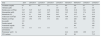
Basal laboratory values during the diagnosis of nephrotic syndrome and in the first outpatient visit after discharge.
| Date | 2014 | 20/3/2015 | 22/3/2015 | 24/3/2015 | 25/3/2015 | 30/3/2015 | 06/4/2015 | 13/4/2015 | 27/4/2015 |
|---|---|---|---|---|---|---|---|---|---|
| Creatinine (mg/dl) | 0.7 | 0.7 | 0.8 | 1.1 | 1.1 | 0.9 | 0.9 | 0.7 | 1.1 |
| Albumin (g/dl) | 4 | 1.9 | 2.6 | 2.8 | 2.5 | 3.6 | |||
| Erythrocytes (×106/μl) | 5.07 | 5.97 | 6.47 | 6.56 | 6.66 | 5.94 | 6.16 | 5.81 | 6.05 |
| Hemoglobin (g/dl) | 16.1 | 18.3 | 19.5 | 19.7 | 20.2 | 18.2 | 18.8 | 17.4 | 18.6 |
| Hematocrit (%) | 47.8 | 51 | 54.9 | 56.9 | 56.2 | 52 | 55.7 | 51 | 55.3 |
| Leukocytes (×103/μl) | 5.49 | 7.07 | 8.50 | 8.35 | 10.70 | 7.96 | 10.11 | 9.85 | 8.98 |
| Platelets (×103/μl) | 133 | 159 | 174 | 174 | 202 | 144 | 208 | 182 | 108 |
| Iron (μ/dl) | 132 | 86 | 179 | ||||||
| Ferritin (ng/ml) | 30 | 362 | 185 | ||||||
| Transferrin (mg/dl) | 279 | 69 | 165 | ||||||
| Systematic urine (proteinuria) | (−) | +++ | +++ | +++ | +++ | +++ | (−) | ||
| Proteinuriaa (g/24h) | 24.4 | 26.96 | 1.90 | 0.17 | |||||
| CCr (m/min) | 85 | 90 | 73.5 |
CCr, creatinine clearance.
All other blood analytical determinations showed: glucose 100mg/dl, urea 72mg/dl, uric acid 7.9mg/dl, cholesterol 439mg/dl, LDL-cholesterol 342mg/dl, HDL cholesterol 75m/dl, triglycerides 112mg/dl, total protein 4.9mg/dl, calcium 7.6mg dl, phosphorus 3.6mg/dl, sodium 127mmol/l, potassium 4.5mmol/l. Blood electrophoresis showed an increase in α2 and β globulins levels, with a percent decrease in albumin without monoclonal peaks. Tumor markers (CEA, CA 19.9, PSA) were within normal limits. Serology for hepatitis B, C and HIV was negative. Immunological study (C-reactive protein, rheumatoid factor, ANA, complement and immunoglobulins A and M) was normal. There was a slight decrease in immunoglobulin G of 730mg/dl (normal: 751–1560). Vitamin B12 and folic acid levels were normal. In urine, the electrophoretic study showed non-selective proteinuria and negative Bence-Jones.
Abdominal ultrasound was difficult to assess due to poor acoustic window: the left kidney had a very difficult viewing while the right had a normal morphology and echogenicity, but there was no dilation of the urinary tract. No findings in the rest of the ultrasound examination. Venous Doppler in lower limb excluded DVT.
We proceeded to perform symptomatic treatment of nephrotic syndrome with intravenous use of 20% albumin and furosemide 120mg, spironolactone 12.5mg, enalapril 5mg/day, atorvastatin 40mg/day along with enoxaparin 60mg/day sc. To determine the cause of nephrotic syndrome, a first renal biopsy was performed on the left kidney without getting sufficient material to establish an etiologic diagnosis (2 sclerotic glomeruli). After the first renal biopsy, empirical treatment was initiated with prednisone 60mg/day. The patient was clinically better, and weight and edema loss started. However, in the new proteinuria control performed seven days after starting prednisone, proteinuria remained above 25g. Given the histological lack of available information, it was decided to repeat a second renal biopsy of the right kidney, finding “6 glomeruli per slice plane without significant alterations, and tubule-interstitium component and vessels without findings”, that is why the patient was diagnosed with minimal change glomerulonephritis. Given these histological findings, the established treatment with prednisone was maintained, and 5 days after the second biopsy, proteinuria was reduced to less than 2g. With clinical and analytical improvement, the patient was discharged.
From the first visit to the ER, polycythemia was detected, with the rest of the series in the normal range. The polycythemia has been present during hospitalization and even on the first visit after discharge. Hematology were consulted, and a blood morphology study was requested (red blood cells: normocytic–normochromic with ovalocytes presence and some poikilocytes, white blood cells series: neutrophils with decreased cytoplasmic granulation; Platelet series: without significant morphological alterations); JAK-2 mutations not detected and exon 12 was negative. By these findings, together with the absence of splenomegaly, the polycythemia was considered spurious. Despite clinical improvement, with remission of proteinuria, polycythemia persisted, that is why it was decided to perform a phlebotomy.
The association of nephrotic syndrome and polycythemia is rare. Less common is minimal change glomerulonephritis, the cause of nephrotic syndrome in patients with polycythemia. Well, we are describing a case of this association in a very elderly patient.
Most cases of nephrotic syndrome and polycythemia documented in the literature correspond to focal segmental glomerulosclerosis, mainly related with polycythemia vera,1–3 and, in some isolated cases, with secondary polycythemia,4 Some cases of idiopathic membranous nephropathy and secondary polycythemia,5,6 are also collected. Regarding the association of minimal-change disease and polycythemia, as in our case, in reviewing the literature we have only found a few casos.7,8 Although nephrotic syndrome occurs in childhood, and both entities are treated with glucocorticoids, it is in the second decade of life, coinciding with a nephrotic relapse when polycythemia is discovered. In our case, the beginning of both the nephrotic syndrome and polycythemia occur simultaneously and in the eighth decade of life.
In the cases of minimal-change disease and/or membranous nephropathy associated with polycythemia reported in the literature, it is established the hypothesis that as a result of renal hypoxia and increased secretion of interleukin-8 by the kidney, an overproduction of erythropoietin would take place, although in some of these cases the levels were normal and in others they were not available. In our case, although we do not have the levels of erythropoietin, having ruled out the presence of primary polycythemia and other secondary causes, perhaps this can be explained by the existence of renal ischemia associated with severe nephrotic syndrome.
Glucocorticoid therapy in known cases of minimal-change disease and polycythemia allows the disappearance of proteinuria and the levels of hemoglobin and hematocrit were maintained within normal range. In our patient, treatment with corticosteroids allowed a complete remission of proteinuria and a normalization of the levels of transferrin, while polycythemia persisted until the first outpatient visit, requiring the completion of a phlebotomy as a treatment for this.
In conclusion, we are describing a rare case of minimal-change disease and secondary polycythemia, with a simultaneous onset of both clinical entities in a very elderly patient. Steroid treatment allowed proteinuria remission and transferrin normalization, but without changes in blood parameters. A phlebotomy was necessary to correct the problem.
Conflicts of interestThe authors have no conflicts of interest to declare.
Please cite this article as: Heras M, Saiz A, Rosado B, Fernández-Reyes MJ, Queizán JA, Callejas R, et al. Asociación de nefropatía por cambios mínimos y policitemia en un paciente muy anciano. Nefrología. 2016;36:67–69.


