


The “U-shaped” tunneling procedure of femoral catheters represents a technical variant that is practiced in various hospitals as an anatomical and functional adaptation in patients with complex venous access, although it is rarely described in the literature. This technique creates a curved subcutaneous path that allows for the exit site to be moved away from the inguinal fold, with potential benefits in terms of fixation, comfort and a reduction in infections being reported.1
The motivation to develop this technique arises from the need to optimize femoral access in obese patients with amputations or unfavorable anatomy. Unlike linear tunneling, the “U” curve increases the contact surface between the catheter and the subcutaneous tissue, thereby decreasing the risks of traction, displacement and skin maceration.2 In our experience, we applied this technique in more than 30 patients, without adverse events related to the curved trajectory being reported; moreover, good tolerance, adequate functioning and a low rate of early infection were observed, although formal comparative studies are needed.
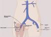
The procedure begins with a detailed ultrasound evaluation, which allows for the estimation of the depth of the femoral vein and the accurate planning of the subcutaneous route, which usually exhibits a length of 15 to 18 cm in the form of a curve (Figs. 1 and 2). Based on these measurements, the appropriate catheter length is selected (usually between 23 and 28 cm), with the aim of achieving a smooth and functional curvature that facilitates tunneling and ensures that the distal tip is correctly located in the inferior vena cava, which is a position that has been associated with a lower incidence of dysfunction and complications.3 This approach allows for the individualization of the choice of a catheter beyond the body mass index (BMI), with consideration of factors such as the distribution of adipose tissue, the length of the extremities and the morphology of the thigh. The curved layout of the trajectory is drawn from configurations that have been previously used for jugular catheters.4


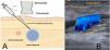
Ultrasound view of the common femoral vein for the planning of femoral vascular access. (A) Schematic representation in a cross section showing the measurement of the skin-femoral vein distance and the orientation of the needle at an angle of 30–45° with respect to the skin, avoiding the femoral artery. (B) Longitudinal ultrasound image with color Doppler of the common femoral vein, in which the skin-vein distance that was used for planning the creation of a “U”-shaped subcutaneous tunnel guided by ultrasound is indicated.
Although some equipment uses curved femoral trajectories, its use is not standardized. Previous studies have demonstrated that the shape of the tunnel influences the rate of femoral infection.4 In addition, other authors have described the practical advantages of the lateral exit site in tunneled femoral catheters.5 Accordingly, our technique strives to transfer these benefits to the femoral terrain through a subcutaneous route in the form of a “U” curve.
We recognize the current limitations of this proposal, which does not include a direct comparison with the straight technique or longitudinal follow-up of complications. However, we believe that its publication as a letter may stimulate other groups to replicate it and evaluate its clinical impact. This communication is aligned with the need to explore innovative strategies in vascular access.6
FinancingThe authors declare that this work has been performed with their own resources.
The authors declare no conflicts of interest.
We are grateful to those researchers who provided their valuable collaboration for the completion of this article.


