To the Editor,
Calciphylaxis is a rare but important cause of morbidity and mortality in chronic kidney failure patients undergoing renal replacement therapy. Its prevalence is increasing and ranges between 1% and 4% in patients undergoing dialysis.1,2 It is characterised by ischaemia and cutaneous necrosis secondary to calcification, fibrodysplasia of the intima and thrombosis of small dermo-epidermic arterioles.1,2
Its pathogenesis is not very well known, although it is associated with different risk factors such as female sex, obesity, diabetes, metabolic syndrome and calcium and phosphorus disorders.3,4 Another factor that may favour this disease is the use of coumarin-based anticoagulant drugs, which favour vascular calcification by means of inhibiting g-carboxylation of vitamin K, depending on the matrix protein Gla (protein that inhibits vascular calcification).1,4
We present the case of a 55-year-old male with a personal history of primary antiphospholipid syndrome with oral anticoagulant agents since 2003, renal clear cell carcinoma. He had a pacemaker because of an atrioventricular block, severe mitral regurgitation and aortic regurgitation, lymph node tuberculosis and operated right hydrocele. In 1993, he was included in a haemodialysis programme due to chronic renal failure of vascular origin. He received three kidney grafts, the last being in 1997, later presenting with thrombosis, for which he started peritoneal dialysis in March 1998. He was transferred to haemodialysis in November 1999, because of a peritonitis-related sepsis caused by Pseudomonas.
He underwent subtotal parathyroidectomy in 2000, due to severe secondary hyperparathyroidism, manifested as elevated parathyroid hormone (PTH) in the biochemical tests that was treated with vitamin D analogues and cinacalcet.
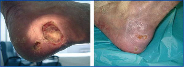
He was admitted in December 2008 for symptoms of heart failure and data showing protein-calorie malnutrition. In the examination, the patient presented with two ulcers on the heel of his left foot. One was approximately 3.5cmx2cm, ulcerated rounded, with red edges, with a necrotic eschar. It was very painful and had preserved distal pulse.
Biochemical tests showed: phosphorus: 4.5mg/dl; corrected calcium: 10.1mg/dl; PTH: 537pg/ml; albumin: 2.2mg/dl; prealbumin: 7.44mg/dl; haemoglobin (Hb) 11.5. Staphylococcus aureus and Clostridium perfringens were found in the cultures. Given that calciphylaxis was suspected, a skin biopsy was performed, confirming diagnosis.
A multidisciplinary approach was adopted: debridement and treatment of the lesions, starting intravenous antibiotic therapy in accordance with the antibiogram. Calcium-based phosphate binders and vitamin D were withdrawn and cinacalcet dose increased. Given the patient's clinical situation (dyspnoea on minimal efforts and poor tolerance to conventional sessions) and with the aim of intensifying the dialysis dose, long nightly haemodialysis was started on a daily basis. Later, to improve calcaemia control, low-calcium dialysis fluid was used (1.75mmol/l). Once the dialysis session had finished, treatment with 25% sodium thiosulphate (ST) at 25g/l/1.73m2 was started.
ST treatment was maintained until the lesion was completely cured, which took 9 months of treatment. The only secondary effect was that the patient presented with nausea and vomiting, but this was resolved with prokinetic and antiemetic agents.
There is no treatment that is specific for calciphylaxis. Special emphasis is currently made to controlling the calcium/phosphate and hyperparathyroidism products, 4,5 suspending treatment with calcium and vitamin D supplements, increasing dialysis frequency to 5 or 6 weekly sessions, reducing the calcium concentration in the dialysis solution to 1.5-1mEq/l and treating the lesions very carefullly.1 Also, measures aimed at improving tissue hypoxia were used, such as hyperbaric oxygen therapy.4,6
Recently, other treatment lines have become important, such as intravenous ST, an antioxidant that is capable of binding to the nitric oxide synthase enzyme and the calcium binder. In the cases described, ST rapidly alleviated pain but resolved ischaemic ulcers slower.1-4, 6, 7 Its antioxidant properties correct endothelial dysfunction and favour vasodilatation. Furthermore, together with calcium it forms calcium thiosulphate, which is more soluble than calcium phosphate (present in vascular calcification), which could help eliminate calcium deposits.1-4, 7 There is no consensus with regards treatment duration but most authors agree that it should be maintained until ulcers have completely healed. Adverse reactions are often mild and includes nausea, vomiting, headaches, nasal discharge and metabolic acidosis.1 Less common reactions are disorientation, hallucinations, arthralgia, cramps or high blood pressure, among others.1
More cases have been reported in the literature, patients treated with biphosphonates whose lesions regressed.4,7-9 Cinacalcet has also proven to be beneficial. However, current treatment continues to present with poor prognosis, with a mortality rate between 60% and 80%, and sepsis is the most common cause of mortality.1-4, 7
Figure 1. Evolution of the lesion


