

Fracture risk assessment in patients with chronic kidney disease (CKD) has been included in the CKD-MBD ("Chronic Kidney Disease-Mineral and Bone Disorders") complex in international and national nephrology guidelines, suggesting for the first time the assessment of bone mineral density (BMD) if the results can influence therapeutic decision-making. However, there is very little information on actual clinical practice in this population. The main objective of the ERCOS (ERC-Osteoporosis) study is to describe the profile of patients with CKD G3-5D with osteoporosis (OP) and/or fragility fractures treated in specialized nephrology, rheumatology and internal medicine clinics in Spain. Fifteen centers participated and 162 patients (mostly women [71.2%] postmenopausal [98.3%]) with a median age of 77 years were included. Mean estimated glomerular filtration rate (eGFR) was 36 mL/min/1.73 m2 and 38% of the included patients were on dialysis. We highlight the high frequency of prevalent fragility fractures [37.7%), mainly vertebral (52.5%) and hip (24.6%)], the disproportionate history of patients with glomerular disease compared to purely nephrological series (corticosteroids) and undertreatment for fracture prevention, especially in nephrology consultations. This study is an immediate call to action with the dissemination of the new, more proactive, clinical guidelines, and underlines the need to standardize a coordinated and multidisciplinary care/therapeutic approach to these patients in an efficient way to avoid current discrepancies and therapeutic nihilism.
La valoración del riesgo de fractura del paciente con enfermedad renal crónica (ERC) ha sido incluida en el complejo CKD-MBD («Chronic Kidney Disease-Mineral and Bone Disorders») en guías nefrológicas internacionales y nacionales, sugiriéndose por primera vez la evaluación de la densidad mineral ósea (DMO) si los resultados pueden condicionar la toma de decisiones terapéuticas. Sin embargo, existe muy poca información en práctica clínica real en esta población. El objetivo principal del estudio ERCOS (ERC-Osteoporosis) es describir el perfil de los pacientes con ERC G 3-5 D con osteoporosis (OP) y/o fracturas por fragilidad atendidos en consultas especializadas de nefrología, reumatología y medicina interna en España. Participaron 15 centros y se incluyeron 162 pacientes (siendo en su mayoría mujeres [71,2%] postmenopáusicas [98,3%]) con una mediana de edad de 77 años. La mediana del filtrado glomerular estimado (FGe) fue de 36 mL/min/1,73m2 y 38% de pacientes incluidos estaban en diálisis. Destacamos la elevada frecuencia de fracturas por fragilidad prevalentes [37,7%), principalmente vertebrales (52,5%) y de cadera (24,6%)], el antecedente desproporcionado de pacientes con patología glomerular en comparación a series puramente nefrológicas (corticoides) y el infratratamiento para la prevención de fracturas, fundamentalmente en consultas nefrológicas. Este estudio supone una inmediata llamada a la acción con la difusión de las nuevas guías clínicas, más proactivas, y subraya la necesidad de homogeneizar el enfoque asistencial/terapéutico multidisciplinar coordinado de estos pacientes de un modo eficiente para evitar las actuales discrepancias y el nihilismo terapéutico.
It is well known, not only in the world of nephrology, that the international organization Kidney Disease Initiative Global Outcomes (KDIGO) defines chronic kidney disease (CKD) as the presence of alterations in renal structure or function, for a period of more than 3 months, with an impact in patient´s health.1–4 Among its complications, one of those with greatest impact is the negative effect on bone health; therefore, the 2009 KDIGO guidelines included the term Chronic Kidney Disease-Mineral and Bone Disorder (CKD-MBD) to describe a systemic entity that integrates all the biochemical, skeletal and extra-skeletal alterations occurring as a consequence of the mineral metabolism disorders of CKD, and which are associated with an increase in cardiovascular disease and mortality.4–6
However, it is much less well known in the nephrology and other more general fields that osteoporosis (OP) is one of the diseases recently considered in the CKD-MBD complex. OP is defined as the loss of global mechanical resistance of the bone that predisposes to an increased risk of fractures.7–9 These fractures have a high morbidity and mortality rate for patients, in addition to a high socioeconomic cost.10,11 Although there is very little information about OP in CKD and its different stages (G), an increased risk in the incidence of fragility fractures has been described in patients with CKD as compared to the general population, and these fractures are associated to an increased mortality.12,13
For the diagnosis of OP, it is useful to verify the existence of a reduction in bone mineral density (BMD) by means of Dual-energy X-ray Absortiometry (DXA). However, in the nephrology field, BMD measurement had never been routinely considered in patients with CKD because it was believed that it did not predict the risk of fracture as in the general population, arguing that the decrease in BMD could be a consequence of the alterations in calcium-phosphorus metabolism associated with CKD.14 However, several studies confirmed not only that there is a direct relationship between the degree of CKD and BMD loss,15–19 but also a clear association between decreased BMD and increased risk of fracture in this population.20 In fact, BMD measurement by DXA is useful to predict fractures across the entire spectrum of CKD G3a-5D.21–24 All this evidence was reflected in the 2017 KDIGO guidelines update,4 where BMD assessment in CKD G3-5D patients with evidence of CKD-MBD and/or risk factors (RF) for fracture was suggested for the first time if its results could impact therapeutic decisions. This suggestion has also been adapted in the recent recommendations of the Spanish Society of Nephrology (S.E.N.) for the management of bone-mineral metabolism disorders in patients with CKD 2021 (S.E.N.-MM).6,14 In addition, the Spanish Society of Rheumatology (SER) has reinforced this position by highlighting patients with CKD as candidates for DXA due to their high risk of OP and fragility fractures.25
However, there is ambiguity in the guidelines that still generates notable discrepancies as to when to perform DXA in patients with CKD. Moreover, DXA is likely to underestimate the risk of fracture in these patients, as it provides information mainly on bone "quantity" but not on bone "quality", which in this population is usually impaired, due to alterations in bone remodeling, mineralization and volume.21,26
There is no doubt that the performance of DXA in patients with CKD could help to improve the prevention of fractures still present in these patients, especially taking into account the growing experience that shows that treatments for OP are also effective in patients with CKD.27 This paradigm shift could probably contribute to reducing the incidence of fractures and their associated morbidity and mortality (primary and secondary prevention), which until now were not considered in the guidelines despite the great health care and economic burden they entail.3,28–30
For all these reasons, the Chronic Kidney Disease and Osteoporosis (CKD and Osteoporosis) study (ERCOS) was designed with the aim of defining the profile of patients with CKD G3-5D (main objective), as well as to know the RFs associated to bone fracture, and the diagnostic and therapeutic strategies (secondary objectives). The results of this study will help us to deepen our knowledge on the relationship between CKD and OP, and to improve the medical care of these patients.
MethodologyDesignERCOS is a descriptive, observational, cross-sectional, multicenter, nationwide, epidemiological study in which data from patients with CKD G3-5D, diagnosed with OP in routine clinical practice, were collected consecutively for 9 months. The study was carried out in nephrology, rheumatology and internal medicine departments of a total of 15 hospitals throughout Spain.
PopulationWe consecutively included adult patients (>18 years) with a diagnosis of CKD G3-5D, with an estimated glomerular filtration rate (eGFR) <60 mL/min/1.73 m2 (including dialysis patients), who also had been diagnosed with OP by DXA (T-score ≤−2.5 in lumbar spine, femoral neck or total hip) and/or with a history of at least one fragility fracture (those caused by low-impact trauma, such as falling from one's own height, mainly those of the vertebra, hip, proximal humerus and distal radius) occurring after the age of 50 years.
Data collectionIn a single visit, after obtaining the patient's informed consent, demographic data, comorbidities, RF of fracture, previous fractures, BMD values, mineral metabolism parameters and treatments received for OP (type and prescriber of the treatment) were collected.
Statistical analysisThe data recorded in the electronic data collection logbook (DCL) of the study were analyzed by descriptive analysis. The mean and standard deviation or median and interquartile range were obtained as appropriate according to the normality distribution of the quantitative variables. For qualitative variables, the frequency and percentage of the valid N were obtained.
To describe and compare variables by renal stage, patients were categorized based on eGFR or need for dialysis. Comparison of study variables by renal stage was performed using Pearson'sχ2 test for qualitative variables and one-factor analysis of variance (ANOVA) for quantitative variables. In case of statistically significant differences in the overall comparisons, post-hoc two-to-two were performed using the Bonferroni test.
For the comparison of BMD, we evaluated whether the change was significantly different from zero using Student's t-test. For the comparison of the prescription of treatments for OP according to medical specialty, variables were compared using Pearson'sχ2 test for qualitative variables and one-factor ANOVA for quantitative variables. All statistical tests were bilateral establishing the significance level α at 5%.
Ethical considerationsApproval was obtained from the Ethics Committee for Drug Research (CEIm) of the Complejo Hospitalario Universitario de Canarias and, additionally, it was also evaluated and positively assessed by the local CEIm of each participating hospital. The standards of good clinical practice and the ethical principles established for research on human subjects in the Declaration of Helsinki and its subsequent revisions were respected. All study participants were informed about the study and voluntarily agreed to participate by signing the informed consent form.
ResultsA total of 162 patients (115 women) were included. CKD G3 (n = 65), CKD G4-5 (n = 35)and CKD G5D(n = 62). The most frequent profile of the patient with CKD and OP in this population was female (71.2%) postmenopausal (98.3%) of advanced age (77 years median), with a BMI of 26.4 kg/m2, a serum creatinine of 1.48 mg/dL (1.20–2.01) and an eGFR of 36 mL/min/1.73 m2 (26–43). The sociodemographic characteristics by stage of renal disease are shown in Table 1. Thirty-eight percent of patients were on dialysis, with a median time on dialysis of 2.6 years (1.3–4.3 years). The most frequent etiology of CKD was arterial hypertension (47.5%), followed by glomerular diseases (26.5%) and diabetes mellitus (19.8%). In 14.2% of cases the cause was unknown. Twenty-one percent of the patients received steroid treatment (10.8%, 20.6% and 32.3% of patients with CKD G3, G4-5 and G5D, respectively) and 71% of these were receiving steroids at the time of inclusion in the study.
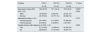
Demographic characteristics and prevalence of fragility fractures by chronic kidney disease stage (CKD-stage).
| Variable | CKD 3 | CKD 4-5 | CKD 5D | P value* |
|---|---|---|---|---|
| N = 65 | N = 35 | N = 62 | ||
| Age (years), mean (SD) | 78.2 (9.76) | 74.1 (12.4) | 70.9 (12.4) | 0.002a |
| Sex, n (%): | 0.200 | |||
| Male | 16 (24.6%) | 8 (22.9%) | 23 (37.1%) | |
| Woman | 49 (75.4%) | 27 (77.1%) | 39 (62.9%) | |
| Menopausal status, n (%): | 0.167 | |||
| Posmenopausal | 49 (100%) | 27 (100%) | 37 (94.9%) | |
| Premenopausal | 0 (0.00%) | 0 (0.00%) | 2 (5.13%) | |
| Fragility fracture after diagnosis of CKD, n (%): | 0.553 | |||
| Yes | 27 (41.5%) | 13 (38.2%) | 20 (32.3%) | |
| No | 38 (58.5%) | 21 (61.8%) | 42 (67.7%) | |
SD: standard deviation; CKD: chronic kidney disease.
Arterial hypertension, diabetes mellitus, cardiovascular disease (including heart failure and ischemic heart disease) were the most frequent comorbidities (87.1, 35.6, 29.4 and 13.5%, respectively). When the prevalence of RFs of fracture was analyzed, diabetes mellitus was identified in more than 30% of patients, hypogonadism or early menopause (9.4%), liver disease (9.4%) and hip fracture in father and/or mother (8%). Diabetes mellitus, hypogonadism or early menopause and liver disease were more frequent in dialysis patients compared to patients with CKD G3 and CKD G4-5 (p = 0.013 and p = 0.021, respectively).
The study showed a prevalence of fragility fractures of 37.7%. A 19.1% had at least one vertebral fracture and 9.3% had a hip fracture.
The most frequent locations of fractures were: vertebral (50.8%), hip (24.6%), wrist (21.3%), humerus (16.4%) and pelvis (13.1%). A total of 19.4% of patients with CKD suffered a fragility fracture despite receiving treatment of OP.
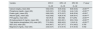
With respect to the biochemical parameters of bone metabolism, dialysis patients as compared to CKD patients not on dialysis (CKD G3-5D), had significantly lower serum levels of calcium, total protein, albumin and 25-hydroxyvitamin D, and higher levels of phosphorus, parathyroid hormone and alkaline phosphatase, (Table 2).
Biochemical data of phosphocalcium-calcium metabolism according to CKD stage.
| Variable | ERC 3 | ERC 4-5 | ERC 5D | P value* |
|---|---|---|---|---|
| N = 65 | N = 35 | N = 62 | ||
| Calcium (mg/dL), mean (SD) | 9.62 (0.50) | 9.45 (0.52) | 8.80 (0.64) | <0.001a,b |
| Phosphorus (mg/dL), mean (SD) | 3.45 (0.61) | 3.71 (0.90) | 4.44 (1.24) | <0.001a,b |
| Protein (g/dL), mean (SD) | 6.89 (0.63) | 6.73 (0.57) | 6.46 (0.54) | <0.001a |
| Albumin (g/dL), mean (SD) | 4.32 (0.82) | 4.26 (0.76) | 3.80 (0.41) | <0.001a,b |
| PTH (pg/mL), mean (SD) | 102 (76.3) | 168 (180) | 317 (276) | <0.001a,b |
| 25-hydroxyvitamin D (ng/mL). mean (SD) | 32.6 (13.8) | 36.0 (20.1) | 23.9 (13.6) | <0.001a,b |
| Total alkaline phosphatase (UI/l), mean (SD) | 77.0 (28.0) | 88.8 (40.0) | 116 (61.9) | <0.001a,b |
| GGT (IU/l), mean (SD) | 34.9 (49.7) | 42.7 (47.7) | 37.4 (40.2) | 0.755 |
| Magnesium (mg/dL), mean (SD) | 2.02 (0.37) | 2.13 (0.73) | 2.15 (0.43) | 0.442 |
CKD, chronic kidney disease; GGT, gamma glutamyl transpeptidase; PTH, parathyroid hormone; SD, standard deviation.
Forty-six percent of patients were receiving calcium supplements and 84.7% were on treatment with vitamin D (calcidiol 58%; cholecalciferol 45.7%), with no significant differences between the different stages of CKD. A total of 23.9% of patients received treatment with paricalcitol (especially in CKD G4-5D) and 8% were on calcitriol. Sixteen percent of patients were on calcimimetics, and 35.8% were receiving phosphate binders (31.4% of patients with CKD G4-5 and 77% of patients G5D; p < 0.001). Of these phosphate binders, 37.9% were calcium-based binders, especially calcium carbonate (50%), and 82.8% were non-calcium-based binders, the most frequent being sevelamer carbonate in 62.5% of patients. A total of 20.7% of patients, all of them except one on dialysis, received both types of binders.
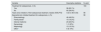
For the diagnosis of OP, a DXA was performed in 157 patients (97%), with a median time of 2.7 years before inclusion in the study (Table 3). Diagnostic DXA was performed much earlier in patients with CKD G3 than in those with other stages (time elapsed from diagnosis of CKD and performance of DXA in patients with CKD G3 vs. CKD G4-5 and CKD5D was 7.5 ± 11.4 vs. 3.4 ± 3.1 and 1.8 ± 1.9 years, respectively; p < 0.001). The mean T-score value was in the range of OP in the lumbar spine, and in the range of osteopenia in hip and femoral neck. Mesurement of BMD in radium, suggested in some guidelines for diagnosis of OP in patients with CKD, was measured in only 16% of patients.
Bone densitometry at the time of diagnosis of osteoporosis/osteopenia.
| Variable | Descriptive statistics | N valid |
|---|---|---|
| T-score lumbar spine, median (P25,P75) | −2.55 (−3.27,−1.83) | 146 |
| T-score hip, median (P25,P75) | −2.30 (−2.70, −1.70) | 143 |
| T-score femoral neck, median (P25,P75) | −2.30 (−2.80, −1.70) | 142 |
| T-score ultradistal radius, medium (P25,P75) | −1.70 (−2.70, −0.30) | 25 |
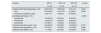
A 60.1% of patients had been on specific treatment for OP for a median of 4 years (P25, P75: 1.7–6.5). This treatment was initiated in 86.2% of patients with CKD G3, 65.7% with CKD G4-5 and 30.6% on dialysis (p < 0,001), 6.3 ± 5.4, 4.9 ± 4.3 and 2.3 ± 2.5 years, respectively before inclusion in the study (p = 0.008). Median eGFR at initiation of OP treatment was 44 mL/min/1.73 m2 (35–54) in patients not on dialysis (CKD G3-5D).
Physicians prescribing treatment for OP were 46.9% rheumatologists, 23.5% internal medicine, 10.2% primary care physicians and 13.3% nephrologists (Table 4). In patients with CKD G5D, prescription was mostly issued by nephrologists (36.8%) and rheumatologists (31.6%) and to a lesser extent by primary care physicians (10%), internal medicine (15.8%), among others.
Treatment for osteoporosis and prescribing physician according to specialty.
| Variable | Descriptive statistics | N valid |
|---|---|---|
| Treatment for osteoporosis, n (%) | 163 | |
| Yes | 98 (60.1%) | |
| No | 65 (39.9%) | |
| Years since initiation of first osteoporosis treatment, median (P25,P75) | 4.02 (1.66–6.54) | 98 |
| Specialist who initiated treatment for osteoporosis, n (%) | 98 | |
| Rheumatology | 46 (46.9%) | |
| Family doctor | 10 (10.2%) | |
| Internal medicine | 23 (23.5%) | |
| Gynecology | 1 (1.02%) | |
| Nephrology | 13 (13.3%) | |
| Othera | 5 (5.10%) |
By CKD stage, drugs for OP were prescribed primarily by rheumatologists (51.8%) and internal medicine (30.4%) in CKD G3 (82.2% overall) and by nephrologists and rheumatologists in CKD G5D (36.8 and 31.6%, respectively). The time period since the initiation of treatment by nephrologists was nominally less than rheumatologists and internists (2.0 ± 2.0 vs. 5.2 ± 5.0 ) and other specialties (7.8 ± 4.9 years) − (p = 0.006).
The most commonly used antiresorptive treatments were bisphosphonates (45.9% of patients), which in 82.2% of cases were given orally, and were more commonly used in patients with CKD G3 (51.8%) (p = 0.053) (Table 5). The most commonly used oral bisphosphonates were alendronate (45.9%) and risedronate (45.9%). A 17.8% of patients received zoledronic acid (7 patients with CKD G3 and one with CKD G4-5). A lower tendency to receive bisphosphonates was observed in patients on dialysis (21.1% of OP treatments in this stage), but not in patients with CKD G4-5 (52.2%) compared to stage G3 (51.8%) (p = 0.053). A 45.9% of patients initiating OP treatment received denosumab, of which 42.9% of patients had CKD G3, 34.8% CKD G4-5, and 68.4% CKD G5D (p = 0.073).
First antiosteoporotic treatment according to stage of CKD.
| Variable | ERC 3 | ERC 4-5 | ERC 5D | p value |
|---|---|---|---|---|
| N = 56 | N = 23 | N = 19 | ||
| Treatment with bisphosphonates, n (%) | 29 (51.8%) | 12 (52.2%) | 4 (21.1%) | 0.053 |
| Oral | 22 (75.9%) | 11 (91.7%) | 4 (100%) | 0.385 |
| Intravenous (zoledronic acid) | 7 (24.1%) | 1 (8.33%) | 0 (0.00%) | |
| Oral bisphosphonates, n (%) | >0.999 | |||
| Alendronate | 10 (45.5%) | 5 (45.5%) | 2 (50.0%) | |
| Risedronate | 10 (45.5%) | 5 (45.5%) | 2 (50.0%) | |
| Ibandronate | 2 (9.09%) | 1 (9.09%) | 0 (0.00%) | |
| Treatment with denosumab, n (%): | 24 (42.9%) | 8 (34.8%) | 13 (68.4%) | 0.073 |
| Treatment with teriparatide, n (%): | 2 (3.57%) | 1 (4.35%) | 2 (10.5%) | 0.382 |
| Treatment with SERM, n (%): | 1 (1.79%) | 2 (8.70%) | 0 (0.00%) | 0.232 |
CKD, chronic kidney disease; SERM, selective estrogen receptor modulator.
Two patients with CKD G3, one patient with CKD G4-5 and 2 patients with CKD G5D had received teriparatide as first treatment for OP, in all cases prescribed by rheumatologists or internists. Only one patient with CKD G3 and 2 patients with CKD G4-5 had received a selective estrogen receptor modulator (SERM) drug as first treatment.
No patient had received romosozumab (not marketed in Spain at the time of data collection).
The median duration of first treatment for osteoporosis was 3 (1–5) years for bisphosphonates, 3 (1–4) years for denosumab, one (1−1) year for teriparatide and 4.5 (3.7–5.2) years for SERMs.
Of the patients who received a first treatment for OP, 25.5% received a second treatment (denosumab 64%, bisphosphonates 32% and teriparatide 4%). The most frequent causes of treatment change were: loss of BMD (32%). A new fragility fracture (20%) and adverse effects (8%). In 2 patients with CKD G3, the reason was discontinuation of denosumab and initiation of zoledronic acid.
DiscussionThe ERCOS study shows the profile of patients with CKD and OP in Spain. They are mostly women, postmenopausal, of advanced age, with varying degrees of renal disease, including many cases of patients on dialysis.
CKD and OP are highly prevalent especially in older patients. Data from recent Spanish studies indicate that up to 37% of patients ≥ 65 years of age may suffer from CKD and that 1/3 women and 1/5 men will suffer a fragility fracture as a complication of osteoporosis.29,31 However, we did not have data reporting the characteristics of patients in whom these 2 diseases coexist, which is a differential factor with other studies. In fact, being a descriptive study under clinical practice conditions, ERCOS may not reflect population data in patients with CKD. In this sense, analyzing the fracture risk factors in our cohort, the prevalence of diabetes would be very similar to the prevalence described by Lee WC et al.32 in patients with CKD G3-5D, but higher than the 13.1% described by Naylor KL et al.22 in patients with GFR < 60 mL/min/1.73 m2 (their only inclusion criterion). As would be expected, in the latter comparison between their patients with GFR > or < to 60 mL/min/1.73 m2, the prevalence of diabetes and HT was higher in patients with decreased GFR (6.6 vs. 13.1% and 32.3 vs. 58.1%, respectively).22 It is obvious that the inclusion of a greater number of patients with advanced CKD in our cohort would (including a significant number of patients with G5D CKD) easily explain our higher prevalence of HT, DM and even liver disease, as well as the higher prevalence of fragility fractures which, in addition and by definition, was a potential criterion for inclusion in ERCOS. We also highlight the low prevalence of hypogonadism (9.4%) described in our cohort, including early menopause, which is well below the high prevalence of early menopause described in women with CKD (26%).33 Regardless of the fact that our cohort includes 29% males, it is likely that hypogonadism was also underreported in nephrological medical records, which also serves as an alert call to this relevant factor that is not frequently considered so far and may potentially grant diagnostic-therapeutic preferences to these patients.
Taking into account these considerations about our inclusion criteria (CKD and OP) and that the diagnosis of a fragility fracture already has per se diagnostic (OP) and therapeutic (secondary prevention) implications, it is not surprising that the prevalence of fractures at the time of diagnosis of OP in our cohort was almost 40%, higher than the cohort of Naylor et al. in patients with FGe < 60 mL/min/1.73 m2 (25.3 vs. 17.1% in patients with FGe > 60 mL/min/1.73 m2)22 which translates that in many patients the diagnosis of bone disease is made too late. As mentioned previously, although there is little and scattered information on OP in the context of CKD and its different stages, an increased and progressive risk in the incidence of fragility fractures has been described with respect to the general population.12,13,22,34 The fact that most of the fractures observed in these patients affects vertebra and hip makes the situation even more worrisome, since these are fractures associated with greater morbidity and mortality, which in turn is further increased in patients with CKD, who already have more comorbidities, greater cardiovascular risk and lower life expectancy.4,13
It is well known that among the multiple complications of CKD, bone abnormalities is one of the greatest impact, so that the CKD-MBD complex has been confirmed as a systemic entity also associated with fractures.4,35 In this regard, it is worth highlighting the important paradigm shift of the latest national and international guidelines in which it is now suggested to measure BMD in patients with CKD G3a-G5D with evidence of CKD-MBD and/or RF of OP.6,14 The nephrology community recognizes that bone fragility associated with CKD is not only secondary to the "renal osteodystrophy" that is being treated with specific drugs such as phosphate binders, native vitamin D and/or active vitamin D derivatives and/or calcimimetics. New evidence led to important changes showing that DXA measurement of BMD does predict the risk of fracture in CKD and that drugs aimed at treating OP in the general population also improve BMD in these patients.4 Thus, in addition to the need to recognize "hidden" CKD and assess the RF associated with classic OP (senile, postmenopausal), its diagnosis and potential treatment with antiosteoporotic agents have become a new unavoidable challenge.13,29 In this regard, the current trend is to adopt proactive attitudes,3,13 even in the absence of the previously recommended bone biopsies,4,6 with the active participation of the patient in decision-making being necessary given the probable positive risk-benefit balance, although the absence of absolute evidence must be recognized.4,6,28 Consequently, knowledge of the most frequent profile from the results of this study would allow more efficient initial screening.
It is necessary to emphasize once again that the presence of one or more fragility fractures is diagnostic of OP.25 Furthermore, not only the risk of hip fracture and non-vertebral fractures is higher in CKD,36 but it has been also described an association between the risk of fracture and the degree of renal function impairment; although there are not many studies that evaluate vertebral fractures in CKD, certainly they do not seem negligible in this population.36–38
In our study, 37.7% of the patients had suffered a fragility fracture, being the vertebral fracture the most frequent, probably, as mentioned above, due to the high proportion of glomerular disease and its treatment with glucocorticoids. Regardless of the location, a history of fracture implies an increased risk of new fractures throughout life, this risk being especially high during the 2 years following the fracture.39 For this reason, it would seem important to highlight the importance of identifying fractures (especially vertebral fractures, which are frequently asymptomatic) in the evaluation of the patient with CKD, particularly in those patients with the aforementioned characteristics. The diagnosis of fragility fracture classifies the patient as being at high or very high risk of fracture and, therefore, these patients are candidates for treatment for OP in secondary prevention.29
Under conditions of routine clinical practice, it has been observed an evident undertreatment of patients with CKD diagnosed with OP. According to the data of the present study, almost 40% of patients remain untreated. Furthermore, it is important to highlight that significant differences have been observed in relation to prescription by clinical specialty. Most of the treatments for OP were initiated by rheumatologists, with undertreatment by nephrologists. Interestingly, this undertreatment is more evident in early stages of CKD, where the indication for its use would not be limited by the presence of a decreased GFR. In addition, it is possible that patients with early stages of CKD were seen by rheumatologists and internists, with a more proactive attitude in the treatment of osteoporosis.
The selection and prescription of these drugs in the ERCOS study is in line with the treatment recommendations of the main current reference clinical practice guidelines. Oral bisphosphonates are recommended first-line antiresorptive agents for fracture prevention.40,41 Regarding safety, alendronate and risedronate are contraindicated, according to the technical data sheet, in patients with creatinine clearance <30−35 ml/min, although some post-hoc studies with few patients have shown that they can also be effective and safe in more advanced stages of CKD. However, recent publications from a UK and Catalonia electronic database report a slightly increased risk of disease progression in patients with G3b-5 CKD treated with oral bisphosphonates,29,42–44 although data from the UK database showed increased survival in treated patients; of note, in this study renal function deterioration/progression was defined by the change in CKD stage which is less precise that the change in renal function.44 We also highlight that, despite the presence of CKD, 17.8% of patients in our cohort received annual zoledronic acid (the vast majority with CKD G3).
Regarding the use of denosumab, although it does not require dose adjustment in patients with CKD, in stages G4-G5D caution is recommended due to the greater risk of hypocalcemia and secondary increase in PTH.29 In addition, adherence to treatment with denosumab is especially important, since its discontinuation produces a rebound effect of bone remodeling, with accelerated loss of BMD and an increased risk of vertebral fractures, sometimes multiple. In this regard, in our cohort, greater adherence was observed with denosumab than with bisphosphonates (84% of the patients continued treatment with denosumab, while only 42% of the patients maintained the treatment with bisphosphonates).
Despite having started treatment for OP, 19.4% of patients with CKD suffered a fragility fracture. It should be remembered that treatment for OP reduces the risk of fracture by 20–70% depending on the drug and the location of the fracture,45 but it does not prevent all the fractures. It should be taken into account that, the patients who were started on treatment likely were at high or very high risk of fracture, and could have had more fractures in the absence of treatment.
Although the present study highlights the scarcity of treatment of patients with CKD and RF of fracture, it should highlighted that one of the main limitations of this study is the small sample size. It should be taken into consideration that the work was initiated in the middle of the COVID-19 pandemic, so that initially a randomization was established prior to the inclusion of patients, but when the restrictions on public health were reduced and the visits were carried out normally, it was decided to add the option of consecutive recruitment. Also,it is necessary to emphasize that our results cannot be extrapolated to the entire population with CKD since our cohort included patients with a diagnosis of densitometric OP and/or fragility fractures, so the prevalence of fractures in our cohort is higher than what we could observe in the overall population of patients with CKD.22 Another limitation is the short treatment time of the patients, which makes it necessary to take the data related to treatment with caution. Finally, there is a bias in the inclusion of dialysis patients, justified by the interest in knowing the diagnostic-therapeutic attitude in these patients. In this sense, analyzing the characteristics of patients in whom these 2 highly prevalent diseases coexist as an inclusion criterion, has allowed us to describe not only the lack of treatment detected but also the low sensitivity on the part of nephrologists when treating OP, despite the important recent changes in our guidelines.4,6 Thus, as mentioned, in patients with CKD G3a-5D with evidence of OP and/or RF of OP, densitometry is now suggested (if the results will impact therapeutic decisions, evidence 2B). In addition, that patients with CKD G1-2 with OP or high risk of fracture are now recommended (evidence 1A) the same treatment as in the general population and that in patients with CKD G3a-3b, with OP or high risk of fracture (and controlled PTH) the same treatment as in the general population is also suggested (evidence 2B).4,6 Various algorithms for action have been published by various societies and authors, including patients with CKD G4-5D,28,29,46 in whom it is highlighted the importance of secondary prevention (in addition to primary prevention) and the need to study metabolic bone disease in patients with CKD, as well as the need to carry out a study of renal function and metabolic bone disease in patients who have suffered a fragility fracture, for which an active search may even be suggested.
ConclusionWe describe, under conditions of routine clinical practice, a high prevalence of fractures and a worrying deficiency of treatment in patients with CKD and OP, which is especially notorious among nephrologists despite the existence of a positive bias determined by the participation in this study of centers and specialists especially interested in this disease.
Defining the poorly known profile of these patients and the clinical approach used in a national cross-sectional multidisciplinary sample has not only made it possible to detect heterogeneity of actions but also to underline the importance of certain patient subgroups, highlight new clinical needs and provide baseline data on which the impact of the new guidelines can be assessed in the future, especially in the field of nephrology.
Considering the paradigm shift of the recent nephrology guidelines, which suggest for the first time the evaluation of BMD in patients with CKD G3-5D, a call to action seems necessary, especially among nephrologists and probably also extensible to the primary care setting.13 In this sense, we consider it necessary to broaden the dissemination of these guidelines,4,6,28,29,46 more proactive in the diagnosis and treatment of OP in CKD, as well as to homogenize the coordinated multidisciplinary care and therapeutic approach to these patients in order to avoid the current discrepancies and therapeutic nihilism.
FundingThis study was funded by Laboratorios Rubió.
Conflict of interestDr. Jordi Bover Sanjuán declares that he has received honoraria for conferences, consultancies and/or travel grants from Abbvie, Amgen, AstraZeneca, Bayer, CSL-Vifor, GSK, Rubió and Sanofi.
Dr. Carlos Gómez declares having received fees for conferences and consultancies from Amgen, Italfármaco, FAES, Gedeon-Richter, Rubió, UCB and Sanofi.
Dr. Enrique Casado declares having received fees for conferences and consultancies from Eli Lilly, Amgen, UCB, Theramex, Italfármaco, Gedeon-Richter, STADA, Bayer, GP-Pharma and Rubió.
Dr. Minerva Rodríguez García states that she has received fees for conferences, consultancies or attendance at courses and congresses from Gedeon-Richter, Rubió, Sanofi, Kyowa-Kirin, Alexion, Takeda, Amgen and Gebro Pharma.
Dr. María Jesús Lloret Cora has received lecture and/or consulting fees from Abbvie, CSL-Vifor, Rubió and Sanofi.
Dr. Cristina Castro states that she has received honoraria for conferences and/or travel grants and congress attendance from Amgen, Novo Nordisk and Boehringer-Lilly.
Dr. Fernando Henríquez-Palop indicates that he has received honoraria as a lecturer from AstraZeneca and Vifor Pharma.
Dr. Laia Gifre declares having received fees for conferences or attendance to courses and congresses from Amgen, UCB, Rubió and STADA.
Dr. Virginia López de la Manzanara declares having received fees for conferences and consultancies from Amgen, Abbott and Fresenius Kabi.
Dr. Ana Laiz states that she has received lecture and consulting fees from Amgen, UCB, Lilly, Abbvie, Pfizer, Novartis, Janssen and MSD.
Dr. Àngels Martinez-Ferrer states that she has received lecture fees and/or travel grants from Eli Lilly, Amgen, UCB, Theramex, Novartis, GEdeon Richter and STADA.
Dr. Vicenç Torregrosa declares that he has no conflict of interest.
Dr. Secundino Cigarrán Guldrís states that he has received lecture fees from Baxter, AstraZeneca, Novo Nordisk, Boeringher, Chiesi and Bayer.
Dr. Jose Luis Górriz declares having received lecture fees from Vifor SCL.
Dr. Marco Montomoli states that he has received lecture and consulting fees from AstraZeneca, Bayer, Baxter and Novonorksik.
Dr. Nayara Panizo declares that she has no conflict of interest.
Dr. Águeda Prior-Español has received lecture fees and/or travel grants from Eli Lilly, Amgen, UCB, Teames and GP-Pharma.
Dr. Ester Costa Moya indicates that she has received congress funding from Rubió, Amgen and Kyowa Kirin.
Dr. Daniel Martínez-Laguna declares having received fees for conferences, consultancies or attendance to courses and congresses from Amgen, Italfármaco, Pierre Fabre, Rubió, Gedeon Ritcher, STADA, Grünenthal, Theramex and UCB.
Dr. Mariano Rodriguez reports having received lecture fees from Amgen, Kyowa, Vifor Pharma, GSK and Rubio.
Dr. Juan Navarro states that he has received lecture and consulting fees from Abbvie, Amgen, Vifor Pharma, Rubió and Sanofi.
The authors would like to thank GOC Health Consulting for support in the development of the study and medical writing support, and Laboratorios Rubió for funding the study.


