To the Editor,
Haemodialysis catheter dysfunction is a complex issue that results in high morbidity rates. Classic studies evaluated the presence of thrombus in catheters using transoesophageal echocardiography.1 However, never has the role of transthoracic echocardiography been studied as a useful tool in the evaluation of haemodialysis catheters. Poorly functioning tunnelled haemodialysis catheters are usually caused by system thrombosis, probably associated with endothelial damage caused by the continuous rubbing of the catheter tip against the vessel wall or right atrium.2 The clinical manifestations of this condition are catheter dysfunction due to lumen obstruction, pulmonary embolism, paradoxical systemic embolism, and system superinfection.3 The majority of cases involve thrombi that develop insidiously.
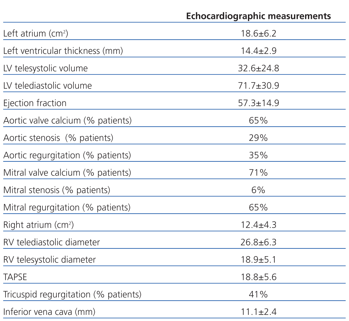
The aim of our study was to evaluate the usefulness of transthoracic echocardiography as an initial diagnostic test for detecting thrombi in patients with tunnelled venous catheters for haemodialysis. Eighteen patients (7 female and 11 male) were monitored at our hospital (mean 39.59±38.12 months on haemodialysis), with a mean age of 72.53±18.09 years. Patient characteristics were: hypertension (82.4%), diabetes mellitus (41.2%), dyslipidaemia (17.6%), tobacco use (23.5%), and previous ischaemic heart disease (11.8%). No patients were diagnosed with thrombophilia; 17.6% were receiving anti-platelets and 5.9% were receiving oral anti-coagulants. All patients underwent transthoracic echocardiograms using a Philips iE33 device. Among the echocardiographic findings, left ventricular hypertrophy with preserved left ventricular systolic function and valve calcification stood out (Table 1). We were able to correctly view the tip of the catheter in 14 cases (77.8%), and only 5 patients underwent catheter replacement due to poor functioning (27.8%).
Thrombi in the right atrium were found in a 36-year-old patient (5.6% of the sample), who was asymptomatic and on haemodialysis due to nephrotic syndrome with focal segmental hyalinosis. These findings were confirmed by transoesophageal examination.
Despite the reduced size of our study sample (the vast majority of which were on chronic dialysis with arteriovenous fistulas), the novelty of this study lies in the fact that it is the first time that a non-invasive, low-cost method such as transthoracic echocardiography has been used to visualise catheters and evaluate the presence of thrombi, so as to avoid potentially severe complications in patients with tunnelled haemodialysis catheters. In our study, we were able to correctly visualise the catheter in a high percentage of patients so we recommend echocardiography on a routine basis in patients with chronic haemodialysis catheters. However, further studies with larger sample size would help reinforce the usefulness of this diagnostic technique.
Conflicts of interest
The authors affirm that they have no conflicts of interest related to the content of this article.
Table 1. Primary echocardiographic findings


