To the Editor,
A dermatological manifestation of chronic kidney disease (CKD) is calcific uraemic arteriolopathy (CUA) or calciphylaxis. It is an anatomopathological entity characterised by necrosis of the skin and adipose tissue due to incorrect calcium salt deposits.1 Morbidity and mortality of calciphylaxis is high due to the complications associated with it: sepsis and ischaemia. Different clinical entities can manifest calciphylaxis: rheumatoid arthritis, inflammatory bowel disease, neoplasias, CKD, systemic lupus erythematosus or HIV infection.1 Its treatment has to be aggressive. Sodium thiosulfate has shown improvements in skin lesions caused by calciphylaxis.
Our patient was a 78-year-old man with history of high blood pressure, diabetes mellitus type 2, dyslipidaemia, CKD of unknown origin treated with haemodialysis three times a week for 3hr, mineral and bone disorder associated with CKD (MBD-CKD), auricular fibrillation and ischaemic heart disease. His usual treatment was sevelamer, enalapril, aspirin, acenocumarol and insulin.
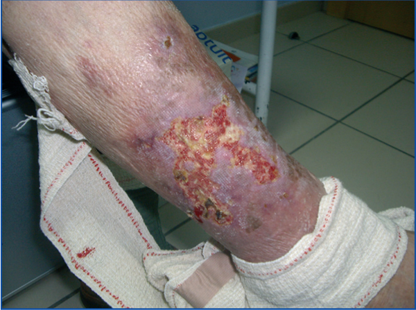
He was admitted to hospital for painful skin erythematous lesions with necrotic edges on both lower limbs, measuring 5x6cm. Physical examination: good general condition, body mass index: 23; blood pressure 150/63mm Hg; heart rate 64bpm; no fever. Cardiopulmonary and abdominal auscultation: painless. Lower limbs: normal pulse, with no sign of deep vein thrombosis and showing previously described lesions. Analyses: parathyroid hormone (PTH): 826.3pg/ml, calcium: 8.9mg/dl; phosphorus; 7.40; creatinine: 9.8mg/dl; albumin: 3g/dl; urea: 156mg/dl; C-reactive protein; 4.3. A cervical ultrasound and parathyroid gammagraphy were performed showing parathyroid hyperplasia free of adenomas. The radiological study (bone series and supra-aortic trunks Doppler) showed vascular calcifications on the ascending and descending aorta. Given the suspected calciphylaxis, we performed a biopsy of one of the lesions, with results compatible with calciphylaxis: lesion with abundant calcium deposits compared with the walls of small vascular structures. Swollen septa due to fibrosis (Figure 1). No signs of necrosis are observed. Lastly, we performed a technetium-99 gammagraphy which did not show that the calciphylaxis spread to the bones.
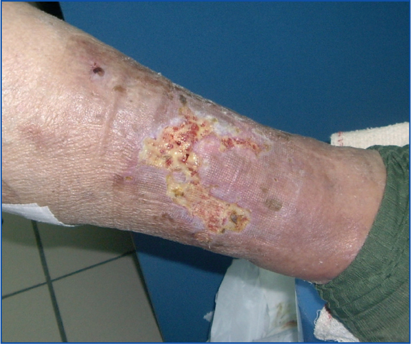
We considered MBD-CKD to be the cause of calciphylaxis and intensified the treatment for it: daily dialysis of 4hr was started with low calcium (2.5mEq/l) in the haemodialysis solution and high flux dialyser. The treatment was intensified with calcium-free phosphate-binders: lanthanum carbonate: 750mg/8hr and sevelamer: 1600mg/8hr, and PTH control with calcimimetics: 60mg/24hr. Acenocoumarol was withdrawn and treatment was started with 80ml of sodium thiosulfate at 25% (20g) following haemodialysis (three times per week). Lesions improved 2 months later (Figure 2). Analytical parameters upon discharge: P: 3.6mg/dl; total Ca: 8.9mg/dl; PTH: 406.90pg/ml.
CUA consists of a hydroxyapatite deposit in the skin and soft tissues with risk of necrosis. In CKD, CUA physiopathology is due to an alteration in phosphocalcic metabolism, uraemic state, increase in PTH (although there are CUA cases following parathyroidectomy)2, calcium-based phosphate binders and a high calcium concentration in the dialysis solution.3 Phosphate and calcium are bound producing vascular, skin and organic calcifications.4
Other predisposing factors are: female sex, obesity,5 hypoalbuminaemia (<2g/dl), diabetes mellitus, C and S protein deficiency,6 oral anticoagulants (they inhibit synthesis of 4.8-gamma-carboxyglumate)7, intravenous iron and vitamin D due to its intestinal action on calcium reabsorption.8
Lesions caused by CUA are often on the abdomen and the calf area, given their abundance of subcutaneous tissue. The lesion is similar to livedo reticularis progressing to ulceration. Calcium deposits in skin are deposited in the dermis and subcutaneous tissue. Physio-pathogenitically, high levels of urea and phosphorus cause smooth muscle cells to convert into osteoblast cells that also increase osteopontin levels, which together with proinflammatory and free radical molecules, make it easier for phosphorus to adhere to calcium.1,7,9,10 This magma is mostly concentrated around calcium deposits that are also found in the arterioles1 and media of the vessels.
The diagnosis is essentially clinical. Lesion biopsy is not recommended given the risk of infection and ulceration.10 Gammagraphy with technetium-99 is used to diagnose whether it has spread to the bones.10 Calciphylaxis has a morbidity and mortality of 80% given the risk of infection and necrosis.
The CUA therapeutic approach must be aggressive, controlling its associated alterations (BMD-CKD control), avoiding agents that could strengthen it, and curing the lesion and infectious complications.2 BMD-CKD control will be performed using calcium-free phosphate binders, low calcium haemodialysis and peritoneal dialysis solutions (CUA can develop above 4mEq/l11) and implementing daily haemodialysis. PTH control will be performed using calcimimetics and vitamin D, preferably vitamin D analogues given that they have a lower calcifying and hyperphosphatemic effect.2 Parathyroidectomy will be reserved for cases resistant to drug treatment.
Dermal CUA lesions that have no ulcerations improve with corticoids.10 However, for those with ulceration, the hyperbaric oxygen chamber is effective against anaerobic organims.11 The sodium thiosulfate (antidote against cynade, used in skin treatments for acne and pityriasis versicolor, and protection against carboplatin and cisplatin toxicity) has proven a successful therapeutic measure in CUA lessions.5,14 Sodium thiosulfate inhibits calcium salt precipitation and dissolves calcium deposits.12,13 It does not have any effects on levels of calcium, phosphorus or PTH. The recommended dose is 20g I.V., three times a week during a minimum of 6 months.2,14 Its administration could lead to metabolic acidosis, osteoclast activation, volume overload and hypotension.
In summary, CUA is a serious entity among our patients. Its treatment has to be aggressive. Sodium thiosulfate is a valid therapeutic agent for treating CUA lesions. It is safe to use and the benefits obtained mean that it can be considered a first-line drug for CUA lesions.
Figure 1. Lesion on right leg prior to treatment with thiosulfate.
Figure 2. Lesion on right leg after two months of treatment with sodium thiosulfate.


