


In Mexico as in the rest of the world there is an increase in the frequency of chronic kidney disease (CKD) that progresses to end stage renal disease (CKD5D) and requires renal replacement therapy such as peritoneal dialysis (PD).1,2 In CKD5D morbidity and mortality are mainly due to cardiovascular disease (CVD).3,4 Uremic cardiomyopathy (UCM) results from abnormalities in cardiac structure and function in patients with CKD3-5. UCM predicts mortality due to the presence of cardiac abnormalities present at the onset of PD4,5 and are characterized by left ventricular hypertrophy (LVH), left ventricular dilatation and both systolic and diastolic heart dysfunction.5 Echocardiography detects abnormalities related to UCM3,4,6 that impact the prognosis of CKD5D patients initiating PD.6 The peritoneal equilibration test (PET)7 evaluates the function of the peritoneal membrane and allows to classify patients as different types of transporters and has a prognostic value.8,9 The aim of the study was to describe echocardiographic findings related to UCM at the commencement of CAPD; these findings were correlated with the type of peritoneal transport (PT).
This is a cross-sectional study carried out from January 2013 to August 2014. We included patients with end stage renal disease of any etiology, between 16 and 75 years, on CAPD for at least one month, with transthoracic echocardiography and PET. We exclude the following patients: with previous diagnosis of heart disease, acute decompensation of any heart disease, dysfunctional peritoneal catheter, peritonitis in the 6 weeks prior to PET, lack of ultrafiltration or acute kidney injury. A transthoracic echocardiogram was performed to determine the ejection fraction (LVEF) and shortening fraction of left ventricle (SFLV). We defined systolic dysfunction (SD) at LVEF values <52% in men and <54% in women, with or without SFLV <25%. We determined left ventricular mass (LVM) and left ventricular hypertrophy (LVH) was defined as LVM >224g in men and >162g in women. To determine the LVH pattern, the relative thickness was calculated with values of <0.42 for eccentric LVH and >0.42 for concentric LVH. We classified as diastolic dysfunction (DD) according to the filling pattern of the left ventricle. Cardiac dimensions and function were calculated according to the recommendations of the American Society of Echocardiography.10 Descriptive statistics, the Kruskall–Wallis test were used to evaluate differences in biochemical or echocardiographic findings in relation to the period (months) on CAPD, as well as Pearson's correlation to determine the relationship between echocardiographic findings and PT.
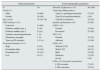
Fifty patients were evaluated, 30 (60%) men, with a median age of 24 years (16–70) and with an average time on CAPD of 2 months (range 1–6). Cause of CKD was unknown in 68% of patients. TP more frequently observed was promedium high in 38% of patients. Biochemically, whole population show moderate anemia and hypoalbuminemia, as well as abnormal fasting glucose. There was LVH in 72%, geometric pattern of concentric type in 53%, pericardial effusion in 32% and pulmonary arterial hypertension in 60%. There was DD in 100% of the patients, mainly with type I pattern or prolonged relaxation in 54% of patients. Fifty percent had Systolic Dysfunction (SD), manifested mainly by decreased LVEF (Table 1). There were valvular calcifications (CV) in 30%; 18% in aortic valve and in 12% in mitral valve. There was no correlation (p=ns) between the different findings related to UCM and the time on CAPD (Table 2). There were no differences in intra- or intergroup comparisons between the PT and the variables analyzed (p=ns). Likewise, there were no correlations of PT with biochemical variables or with echocardiographic findings related to UCM (p=ns).
Demographic, clinical, biochemical and echocardiographic characteristics.
| Clinical parameters | Echocardiographic parameters | ||
|---|---|---|---|
| N | 50 | Diastolic dysfunction n (%) | 50 (100) |
| Gender % | Ventricular filling pattern | ||
| Male | 30 (60) | Type I or prolonged relaxation | 27 (54) |
| Female | 20 (40) | Type II or pseudonormalized | 4 (8) |
| Age (years) | 24 (16–70) | Type III or restrictive | 19 (38) |
| CKD etiology n (%) | Systolic dysfunction n (%) | 25 (50) | |
| Unknown | 34 (68) | LVEF n (%) | |
| Diabetes mellitus type 1 | 5 (10) | Normal | 25 (50) |
| Diabetes mellitus type 2 | 9 (18) | Decreased | 25 (50) |
| Systemic arterial hypertension | 1 (2) | SFLV (%) | |
| Obstructive uropathy | 1 (2) | Normal | 35 (70) |
| Time on PD (months) | 2 (1–6) | Affected | 15 (30) |
| Peritoneal transport n (%) | LVH n (%) | ||
| High | 9 (18) | Without LVH | 14 (28) |
| Promedium high | 19 (38) | With LVH | 36 (72) |
| Promedium low | 16 (32) | Mild | 24 (48) |
| Low | 6 (12) | Moderate | 12 (24) |
| Severe | 0 (0) | ||
| LVH n (%) Geometric parameters | |||
| LVH excentric | 17 (47) | ||
| LVH concentric | 19 (53) | ||
| Biochemical parameters | |||
|---|---|---|---|
| Hemoglobin (g/l) | 8.76±1.4 | ||
| Creatinine (mg/dl) | 10.2±3.9 | ||
| Urea (mg/dl) | 90.9±18.8 | ||
| Glucose (mg/dl) | 108.0±28.7 | ||
| Potassium (mmol/l) | 4.8±0.67 | ||
| Sodium (mmol/l) | 133.1±2.61 | ||
| Albumin (g/l) | 2.9±0.64 | ||
| Phosphorus (mg/dl) | 4.81±1.6 | ||
Measured variables of the patients included are shown. The biochemical parameters and the distribution of the frequencies and the type of PT are described. The echocardiographic parameters are also shown. Results expressed in mean±standard deviation and/or median and range as required (quantitative variables) or numbers and percentages (qualitative variables).
PD: peritoneal dialysis; CKD: chronic kidney disease; SFLV: shortening fraction of left ventricular; LVEF: left ventricular ejection fraction; LVH: left ventricular hypertrophy; n: number of patients.
Comparative and correlation analysis of biochemical variables, echocardiographic findings of UCM with PT.
| Comparison of biochemical variables and echocardiographic findings with PT | Correlation of UCM findings with PT | |||||
|---|---|---|---|---|---|---|
| Variable | p | Finding | p | Finding | r | p |
| Months in PD | 0.72 | Left ventricular ejection fraction | 0.72 | Left ventricular ejection fraction | 0.02 | 0.90 |
| Age | 0.57 | Shortening fraction of left ventricle | 0.35 | Shortening fraction of left ventricle | −0.06 | 0.70 |
| Hemoglobin | 0.50 | Left ventricular mass | 0.85 | Left ventricular mass | 0.1 | 0.50 |
| Creatinine | 0.18 | Relative thickness | 0.44 | Relative thickness | 0.12 | 0.41 |
| Urea | 0.42 | Interventricular septum thickness | 0.97 | Interventricular septum thickness | 0.03 | 0.85 |
| Glucose | 0.11 | Posterior wall thickness | 0.84 | Posterior wall thickness | −0.07 | 0.62 |
| Potassium | 1.00 | End systolic left ventricle diameter | 0.74 | End systolic left ventricle diameter | 0.06 | 0.52 |
| Sodium | 0.39 | End diastolic left ventricle diameter | 0.79 | End diastolic left ventricle diameter | 0.09 | 0.52 |
| Albumin | 0.24 | Left atrium | 0.87 | Left atrium | 0.10 | 0.50 |
| Phosphorus | 0.20 | Aortic root | 0.74 | Aortic root | 0.15 | 0.30 |
| Right ventricle | 0.69 | Right ventricle | −0.02 | 0.89 | ||
| Maximum velocity aorta | 0.47 | Maximum velocity aorta | 0.04 | 0.76 | ||
| Systolic pulmonary pressure | 0.70 | Systolic pulmonary pressure | 0.05 | 0.73 | ||
| E wave | 0.76 | E wave | 0.01 | 0.96 | ||
| A wave | 0.63 | A wave | 0.05 | 0.70 | ||
| Ration E/A waves | 0.48 | Ration E/A waves | 0.06 | 0.70 | ||
Comparison (Kruskall–Wallis test). No statistical difference was observed in biochemical data and echocardiographic findings related to UCM (uremic cardiomyopathy) in relation to PT at the initiation of CAPD. There was not significant correlation (Pearson's r) between TP and echocardiographic findings related to UCM at the start of dialysis.
We describe important findings of UMC present in a young and uninsured population with CKD5D of unknown etiology, since the commencement of a PD program. We also detected hypoalbuminemia and anemia, important factors involved in cardiovascular disease.4 It is important to note that biochemical and echocardiographic alterations were not changed in relation to the months of PD. There was left ventricular dysfunction with DD in all patients, mainly the type of prolonged relaxation. Half of the patients presented SD with frequent LVH, the main manifestation described in the UCM.5 In addition there was vascular calcification present in one third of the patients, mainly in the aortic valve. No kind of PT at the beginning of PD did not correlate with biochemical variables nor with echocardiographic findings related to UCM.
In conclusion, patients with ESRD starting PD show the findings of UCM with predominance of DD, frequent LVH, mainly concentric type and abnormal systolic function. These abnormalities do not change in relation to the time on PD. These patients have important factors described as risk factors for CVD such as anemia, hypoalbuminemia and vascular calcifications. All these abnormalities affect the prognosis of patients with ESRD who initiate PD. However, longitudinal studies are needed to know the evolution of UCM-related findings in patients with PD.
FundingThe study was funded by the Guadalajara Civil Hospital Dr. Juan I. Menchaca.
Conflicts of interestThe authors have no conflicts of interest to declare.
Please cite this article as: Pazarin-Villaseñor L, Reyes-Lopez U, de León-Flores AM, Miranda-Díaz AG, Andrade-Sierra J. Cardiomiopatía urémica y transporte peritoneal en pacientes incidentes con diálisis peritoneal en el occidente de México. Nefrologia. 2017;37:541–544.


