
INTRODUCTION
One of the most common complications in peritoneal dialysis (PD) is poor catheter functioning, which obviously determines the dialysis treatment.
The manifestations of this condition tend to be quite clear: the PD fluid enters and/or exits only with difficulty.
The primary causes of poor catheter functioning include fibrin clot obstruction, catheter tip displacement, and bowel or omental entrapment.
Currently, we have no easily performed imaging tests that can aid in diagnosing the mechanical issues derived from poor catheter functioning. One very simple technique with high value is catheterography. This procedure was first described in the 1990s, but it is used quite infrequently, probably due to the issues of accessibility to radiological rooms.
In our hospital, this procedure has been used for approximately 2 years, thanks to the cooperation of the radiology department. We present below our experience with the technique.
MATERIAL AND METHOD
The procedure takes place in the interventional radiology unit, with the assistance of a radiologist, a nephrologist, and a nurse.
With the patient lying down, the catheter is cleaned with approximately 20ml of saline solution and then 10ml of iodated contrast is infused, under aseptic conditions as always. During the contrast infusion, serial images are digitally recorded, allowing for visualisation of the position of the catheter and of its permeability. The view can be projected in several different formats (antero-posterior, oblique, lateral, etc.).
Whenever necessary, a guide through the catheter can be introduced and relocation can be attempted under direct visual control. In entrapped catheters, this operation is impossible, which indirectly also informs us as to the situation of the catheter.
Once this process is finalised, the catheter is cleaned again and, once back in the peritoneal dialysis unit, intraperitoneal antibiotics are administered as a prophylaxis, and catheter functioning is checked. The patient may return home after the test.
REPRESENTATIVE CASES
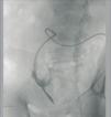
Case 1. 56-year-old; 4 months on CAPD without issues. Episode of severe peritonitis due to an anaerobic bacterial infection. Once recovered, the patient was started on APD. After several weeks, the patient complains of not being able to sleep due to multiple alarms. The treatment regime is evaluated, and a major drainage difficulty is revealed. A simple abdominal x-ray reveals no important findings; the tip of the catheter is observed in the pelvis. A catheterography is administered, which reveals a large loop in the catheter. The situation is resolved through the use of a guide and increased laxative treatment (Figure 1).
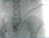
Case 2. 65-year-old woman who started PD after a second transplant. During surgery for the catheter placement, multiple intestinal adherences are freed. From the start, infusion went without difficulty, but drainage was impossible. A catheterography confirmed the suspicion of entrapment; the catheter was then removed through surgery (Figure 2).
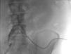
Case 3. 72-year-old patient, who after 15 months on APD started to have issues with drainage, despite laxative use. The catheter tip was found in the epigastrium, and was relocated using a guide (Figure 3).
In our experience, we have not encountered any type of complication: no perforations, haemorrhages, or infections have been produced.
CONCLUSIONS
Catheterography is a safe, easy, and non-invasive method of substantial utility in the differential diagnosis of poor catheter functioning. It can be repeated as many times as necessary, since it presents no risk to the patient.
Conflicts of interest
The authors have no conflicts of interest to declare.

Figure 1. Case 1

Figure 2. Case 2

Figure 3. Case 3


