


Acute Kidney Injury (AKI) is a clinical syndrome characterized by an abrupt decrease in the glomerular filtration rate (GFR) over a short period of time (hours or days). It can result from multiple etiologies, and its common presentation is an increase in serum levels of nitrogenous waste products, which may or may not be accompanied by a decrease in urine output (in two-thirds of cases). In this document, we use the term acute kidney injury (AKI), in accordance with KDIGO.
The syndromic concept of AKI is well-defined, and its detection is based on increases in serum creatinine (SCr). However, over the years, there has been significant disparity in establishing precise defining criteria.1Bellomo et al. proposed the first classification, known as the RIFLE system. Although this classification provided numerous advantages, certain shortcomings became evident over time, such as the underdiagnosis of AKI and the inclusion of estimated GFR (eGFR) in the criteria. Since the estimation of GFR is not valid in acute processes, it was removed from the initial version. For these reasons, the Acute Kidney Injury Network (AKIN) classification was developed in 2007. It is a modification of the RIFLE system that adds an absolute increase in SCr of ≥0.3 mg/dL within a 48-h interval (a cutoff value associated with increased mortality). In 2012, the National Kidney Foundation, through the Kidney Disease: Improving Global Outcomes (KDIGO) workgroup, published the third consensus on the definition and classification of AKI. This new classification merges criteria from both RIFLE and AKIN and is the one currently recommended for use.2
All these classifications are functional ones that allow for the diagnosis and severity staging of AKI. Nevertheless, they have significant limitations, the main one being the use of SCr as the parameter to assess renal function. SCr is known to be a suboptimal marker as it can be influenced by numerous factors such as muscle mass—an important aspect in cases like burn patients who experience muscle loss. Furthermore, previous classifications use the baseline SCr value for diagnosis (defined as the highest value in the last 3 months), which is often unknown. KDIGO guidelines suggest using the lowest SCr value during hospitalization or the value corresponding to a GFR ≥ 75 mL/min/1.73 m² for patients with no prior information.
Using SCr as a marker for AKI can lead to a diagnostic delay since its elevation usually occurs after the drop in GFR has taken place. For this reason, efforts are being made to incorporate biomarkers that allow for the detection of tubular damage, which typically precedes the fall in GFR (see section 2.1).
The incidence of AKI in the general population is estimated at 8.3% (community-acquired AKI), increasing to 21.6% in hospitalized patients and up to 57% in critically ill patients.3,4 Given the lack of a universal definition, mortality rates vary widely from 30% to 67% in critically ill patients, being higher among those requiring renal replacement therapy (RRT)³. Approximately 5%–15% of patients who develop AKI require RRT, though literature values vary depending on the clinical scenario.5 To improve early detection, alert systems can be implemented in the diagnostic process, either as part of hospital information systems or clinical decision-support systems.
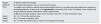
Acute kidney injury risk predictionInjury and risk biomarkersDue to the inherent limitations of SCr, special interest has been focused on new biomarkers that allow for earlier detection of AKI with improved sensitivity and specificity.
Various biomarkers in both blood and urine have been identified that could be useful for early AKI detection, severity stratification, and prognostic assessment. The most promising biomarkers to date include: Kidney Injury Molecule 1 (KIM-1), Neutrophil Gelatinase-Associated Lipocalin (NGAL), Liver-Fatty Acid Binding Protein (L-FABP), Cystatin C, hemojuvelin, N-Acetyl-Glucosaminidase (NAG), netrin-1, gamma-glutamyl transferase (GGT), glutathione S-transferase (GST), Tissue inhibitor of metalloproteinases-2 (TIMP-2), and Insulin-like growth factor-binding protein 7 (IGFBP-7), among others. These molecules participate in different physiological processes altered during AKI, such as renal parenchyma cell death, inflammatory processes, and increased oxidative stress.
Most of these biomarkers are not currently used for AKI diagnosis in routine clinical practice. These biomarkers denote kidney injury, unlike SCr, which denotes a reduction in kidney function. In at-risk patients, the elevation of biomarkers usually occurs before the increase in SCr. Furthermore, cases may occur where biomarkers increase without a corresponding rise in SCr. This situation defines what is known as subclinical AKI. The importance of subclinical AKI is that it may be associated with a worse prognosis.6 Although KDIGO guidelines have suggested that biomarkers could provide added value to SCr determination—thereby improving early AKI diagnosis and prognosis—they have not yet been implemented in clinical practice. Numerous studies indicate these biomarkers could be used in combination with clinical markers to improve risk prediction. In this regard, since 2012, the U.S. Food and Drug Administration (FDA) has approved the use of the TIMP-2/IGFBP-7 ratio as a biomarker for AKI risk stratification, and it has been recommended in clinical guidelines for cardiac surgery.7 In addition to TIMP-2 and IGFBP-7, other biomarkers such as Cystatin C, hemojuvelin, NGAL, and KIM-18,9 might also be useful in estimating the risk of AKI progression. However, this association is not as clear, as contradictory studies exist or suggest that the relevance of these other biomarkers is limited.10 This disparity in results is related to the high heterogeneity of AKI types, the specific AKI definition used, and the sample size of the studies. Therefore, while great progress has been made in validating and applying new biomarkers for AKI diagnosis and prognosis, further research is needed to improve AKI diagnostics. Because current diagnosis remains based on SCr and not on biomarkers, the term AKI is used rather than “Acute Kidney Lesion”.
- •
New biomarkers exist that are associated with the onset and progression of AKI.
- •
These new biomarkers could provide additional value to SCr determination for the early diagnosis of AKI.
- •
Furthermore, they could be useful for stratifying the severity of AKI and evaluating its prognosis.
- •
However, they have not yet been implemented in routine clinical practice.
AKI is one of the most serious and frequent complications in hospitalized patients, entailing high costs and poor global outcomes. Despite its prevalence, impact, and the fact that it is potentially preventable, the onset of AKI often goes unnoticed and is frequently not even recorded in clinical histories or discharge reports. Recognizing the importance of early diagnosis to implement actions aimed at minimizing kidney injury, the introduction of electronic alert systems (e-alerts) within medical record systems has been evaluated in recent years as part of routine clinical practice. The actual effectiveness of these alerts depends on a combination of patient-specific factors, the underlying disease and type of kidney injury developed, the clinical setting, and, above all, the intervention triggered by this early diagnosis.
Perhaps the most representative reports on the utility of these e-alerts are those from the United Kingdom. At the beginning of the last decade, an AKI electronic alert system was mandatorily implemented in inpatient care within the National Health Service (NHS)-UK, which was subsequently extended to Primary Care in a planned manner. These e-alerts are based on the algorithm described in the National Institute for Health and Care Excellence (NICE) guidelines and define the severity of AKI using the KDIGO classification. Laboratory systems automatically calculate the AKI stage based on SCr levels, using the patient's results from the previous year as the baseline creatinine.
The e-alert itself, in addition to identifying the patient, directs the physician toward a decision-support manual. Under this philosophy, each institution opted for different design and implementation modalities for their e-alerts, resulting in significant variability in both the reporting method (email, pop-up windows in the patient's electronic health record, mobile text messages, etc.) and the various clinical action algorithms. Numerous publications have analyzed the impact following their implementation in the United Kingdom, yielding contradictory results. Consequently, in the absence of careful studies regarding both their efficacy and potential adverse effects, various opinions have emerged suggesting the need to moderate the enthusiasm for this type of e-alert.11
In most published studies, no significant impact has been demonstrated regarding short-term mortality or the requirement for dialysis. Nonetheless, positive results have been found concerning a decrease in the prevalence of hospital-acquired AKI, improvements in clinical management, and a reduction in the mean length of hospital stay.12 Results from recent meta-analyses show a high degree of variability in the design of e-alert systems and indicate that the e-alertper se does not improve outcomes unless it is associated with complementary care measures. In such cases, improvements translate into a shorter time for the modification of nephrotoxic drugs, a more rational application of fluid therapy, diuretics, or vasopressors, and more frequent nephrology consultations; this reduces the rate of severe AKI and increases the proportion of patients who achieve renal function recovery.13,14 Recently, results from a large hospital in Birmingham (UK) have demonstrated that, after two years of follow-up post-implementation of e-alerts, the progression of AKI has decreased, and therefore, it is likely that long-term survival will improve. Readmissions to emergency departments following hospital discharge also decreased, a fact the authors attribute to the reduced use of nephrotoxic agents in these patients. The authors emphasize that even minimal changes in patient management can have significant repercussions on long-term outcomes.15
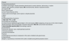
More recently, the NHS has implemented AKI alerts in Primary Care. These experiences have reported an increase in community AKI detection, improvements in follow-up, shorter times to hospital admission, and higher rates of renal injury recovery. The pros and cons of using e-alerts are summarized in Table 1.12,13,15–19
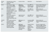
Benefits and limitations of the implementation of e-alerts.
| Benefits | Limitations |
|---|---|
| • Early detection of AKI, providing intervention opportunities • Models based on the increase in SCr are cost-effective and easily implementable • Complex models can facilitate decision-making with an impact on and improvement in patient care. • Can optimize the management of these patients and improve clinical outcomes • Potential utility as quality of care measures | • Dynamic and continuous nature of AKI • If only informative, they have a low impact on patient care • Based on SCr, which is a late marker of kidney damage • Alarm fatigue: alerting on low imminent risk can generate fatigue in clinical teams • Ensuring alert accuracy: false positives and false negatives, applicability • Difficult interpretation of published studies: lack of control or analysis of end events while ignoring the management of less severe cases • The lack of standardization may justify the variability of results obtained between different studies. |
SCr: serum creatinine; AKI: acute kidney injury.
At the consensus conference of the Acute Disease Quality Initiative (ADQI) in 2015, AKI was recognized as an ideal disease state for the application of machine learning and big data. Since then, artificial intelligence has been used to develop AKI risk scales that allow for the implementation of measures in patients at risk of AKI or with early-stage kidney injury. These Machine Learning (ML) models automatically include many variables and allow for the identification of patients at higher risk of adverse outcomes and the discrimination of different kidney injury subgroups. Models published in different AKI settings lack external validation; therefore, the results are not generalizable to other populations. Furthermore, they predict the risk of AKI at a single point in time rather than continuously. On the other hand, there is significant variability among the analyzed cohorts, which, in most cases, are retrospective. Consequently, while the predictive potential of machine learning algorithms is recognized, they still require improvement. Additionally, these models have demonstrated the ability to predict AKI, but not to prevent its occurrence.18,19
- •
Studies on AKI alert systems and clinical decision support continue to demonstrate variable results, which are likely due to differences in local context and implementation strategies.
- •
Further research is required to overcome the validation and implementation barriers of ML models for AKI care.
- •
However, electronic alerts provide the benefits of detection and data collection. In the future, the incorporation of new markers and ML models may make it feasible to "avoid serious consequences of AKI by using these new tools".
One of the key points in AKI management is maintaining an appropriate hydration status. Currently, a wide variety of solutions are available for volume replacement. However, few studies exist, most of them conducted in the critically ill patient setting, that allow for an evaluation of which of these solutions is the most suitable.
The objective of fluid therapy in critically ill patients, and especially in those with septic shock, is to increase preload in order to augment cardiac output. The challenge lies in maintaining adequate tissue perfusion without leading to overhydration. A weight gain exceeding 10% from baseline has been shown to increase mortality in critically ill patients and could have a deleterious effect on renal function.20
In AKI, whether hospital-acquired or community-acquired, one of the main therapeutic pillars is to optimize correct volumetric resuscitation. Resuscitation likely requires individualization in each case, guided by point-of-care ultrasonography (POCUS), capillary refill assessment, or lactate levels.21 Its use should be continuously reassessed to avoid unnecessary fluid overload.
It must be taken into account that fluid therapy is a pharmacological therapy and, as such, can have deleterious effects. There is no ideal composition among the different types of intravenous fluids used.
Three types of fluids are available: colloids, crystalloids, and blood products. The latter are indicated for hemorrhagic shock and will not be discussed in this document.
ColloidsWithin this category, we distinguish between semisynthetic colloids and albumin. The use of semisynthetic colloids was based on maintaining volumetric expansion for a longer duration with a lower volume load. However, in reality, this effect is lost in septic patients due to an increase in endothelial permeability and a higher probability of acute kidney injury (AKI). For this reason, the Surviving Sepsis Campaign guidelines discourage their use, a position endorsed by regulatory agencies such as the FDA and the European Medicines Agency (EMA).22,23 Although the evidence is not as clear, the use of gelatins or dextrans is likewise discouraged.
Regarding the use of albumin as a colloid, it has been shown to be safe in septic patients with respect to the development of AKI, both in the SAFE study and the ALBIOS study, although it should be avoided in patients with traumatic brain injury.24,25
CrystalloidsWithin this category, we find 0.9% saline and balanced solutions. The use of large volumes of 0.9% saline (>2 L) is associated with the development of hyperchloremic acidosis, which negatively impacts the glomerular filtration rate. It is linked to vasoconstriction and a decrease in renal blood flow that could predispose to the onset of AKI.26 In an effort to avoid this effect, balanced solutions have lower chloride concentrations and lower osmolarity. Additionally, they utilize lactate or acetate as a buffer. Although the evidence is weak, there appears to be a benefit to using balanced solutions.27,28 The vast majority of studies provide contradictory results; only the SMART trial showed a positive outcome in renal events at 30 days in favor of balanced solutions.21
- •
Fluid resuscitation must be aimed at increasing cardiac output while avoiding overhydration; therefore, it must be closely monitored (e.g., via ultrasound control, capillary refill time).
- •
In large resuscitation volumes, balanced solutions appear to be safer. The use of albumin has been shown to be safe as a volume expander in septic patients.
Diuretic treatment is indicated in patients with hypervolemia. This clinical situation is common in various pathologies where, in addition, a certain degree of AKI often coexists, such as in heart failure (HF), hepatic failure, nephrotic syndrome, or in septic patients. The use of diuretics in the context of critically ill patients remains a subject of debate. Nevertheless, volume status must be closely monitored. Thus, their use—especially loop diuretics—is particularly indicated in situations involving hypervolemia.25,29
Studies conducted on the effect of loop diuretics as a treatment for AKI have yielded controversial results. Although they do not shorten the duration of AKI or impact mortality, they do decrease the duration of oliguria and the need for renal replacement therapy (RRT).30 There are different classes of diuretics depending on their site of action. The choice of type and dosage will depend on the cause of the AKI, its severity, and the accompanying electrolyte and acid-base disturbances. Since there are few randomized studies on the use of diuretics, treatment must be individualized for each clinical situation.25
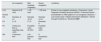
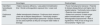
When initiating treatment, it is recommended to assess potential causes of resistance to standard doses. The most common causes of resistance are described in Table 2. Increasing the dose, using the intravenous route, and concomitant use with albumin can help increase the availability of the drug at the tubular level.
Factors for diuretic resistance.31–33
| • Non-hyperhydration (venous stasis, lymphedema) |
| • Hypoalbuminemia |
| • Elevated urea levels |
| • Decreased absorption (intestinal edema in hypervolemic patients) |
| • Decreased renal blood flow and effective circulating volume |
| • Decreased tubular transport (with the concomitant use of NSAIDs) |
| • Decreased renal mass |
| • Activation of the RAAS (renin-angiotensin-aldosterone system) |
| • Hypochloremia |
NSAIDs: non-steroidal anti-inflammatory drugs.
Diuretic types are classified by their site of action:
Loop diuretics. Probably the most commonly used in different types of AKI. They are the most potent diuretics. By blocking the sodium-potassium-chloride cotransporter, they prevent the reabsorption of 20%–25% of the sodium reabsorbed in the renal tubule. They can induce hypotension, primarily due to hypovolemia, but also via vasodilation. Other effects, such as hypokalemia and metabolic alkalosis, occur through the activation of the renin-angiotensin-aldosterone system (RAAS). In rare cases, ototoxicity can occur at high doses, typically with the concomitant use of aminoglycosides.
Chawla et al. standardized the furosemide stress test in critically ill patients, which allows for the assessment of tubular function and predicts progression to more severe kidney injury requiring renal replacement therapy (RRT). Thus, the use of loop diuretics would be recommended for volume control in overhydrated patients who show an adequate response to the stress test.34
Assessing urinary sodium in a spot sample is recommended. Values below 50 mmol/L indicate diuretic resistance in patients with HF and predict a negative sodium balance deficit. Furthermore, hypochloremia is associated with a worse diuretic response in acute HF.This information can help in adopting strategies such as the concomitant use of albumin, 0.9% saline, or hypertonic saline, or combination with other diuretics for sequential nephron blockade.35
In patients with HF and an ejection fraction below 40%, the use of hypertonic saline together with furosemide is associated with an increase in daily urine output, improvement in renal function, decreased hospital stay, and a lower rate of readmission for HF.36
Thiazides. They act by blocking the sodium-chloride transporter in the distal tubule, where 5%–10% of sodium is reabsorbed. Their diuretic effect alone is poor. Secondary effects include hypokalemia and metabolic alkalosis, with a higher rate of hyponatremia. It should be noted that they increase calcium reabsorption and decrease magnesium levels. Their use can increase urine volume in situations of HF refractory to loop diuretics, as part of sequential nephron blockade.37
Potassium-sparing diuretics. Two types of diuretics are distinguished within this group: epithelial sodium channel blockers (amiloride and triamterene) and mineralocorticoid receptor antagonists (spironolactone, eplerenone, canrenone). Unlike all others, the site of action for the latter is at the basocellular level in the distal tubule. The most limiting effect is hyperkalemia. Regarding the former, another effect is their ability to reabsorb magnesium and calcium. Their use is common in situations involving hypervolemia that are refractory to loop diuretic treatment, forming part of the sequential nephron blockade strategy. Such situations are encountered in nephrotic syndrome, patients with liver failure, or HF. However, the use of high doses of spironolactone (100 mg/day) is not associated with an increase in diuresis in patients with acute HF.38 In this latter group of patients, a recent study showed that finerenone resulted in a lower rate of hospitalizations due to worsening HF.39
Carbonic anhydrase inhibitors. This type of diuretic inhibits sodium reabsorption at the proximal level but lacks potent diuretic power. By acting in the proximal tubule, they inhibit the reabsorption of calcium, magnesium, and bicarbonate. Their use is associated with the risk of calcium phosphate lithiasis formation due to increased hypercalciuria and urine alkalization. The recently published ADVOR study demonstrated a reduction in congestion symptoms as well as a decrease in hospital stay when adding 500 mg of intravenous acetazolamide to furosemide treatment.40 It should be noted that, by inducing bicarbonate loss, they can compensate for the alkalosis produced by loop diuretics. Nevertheless, close monitoring is required, as prolonged use can lead to metabolic acidosis.
- •
The use of diuretics is recommended in patients with AKI and overhydration.
- •
Loop diuretics are the first-line indication. Nonetheless, the choice of diuretic type must be individualized. The cause of AKI, as well as any accompanying electrolyte and acid-base balance disturbances, must be taken into account.
Currently, there is no standardized method for dosing medications in AKI, as calculations of creatinine clearance (CrCl) using the Cockcroft-Gault equation or the estimated glomerular filtration rate (eGFR) according to the Chronic Kidney Disease Epidemiology Collaboration (CKD-EPI) are not valid when SCr is not stable. During the development of AKI, the estimated CrCl (by Cockcroft-Gault) or eGFR overestimates renal function and can lead to drug accumulation and potential toxicity; during the recovery phase, they underestimate renal function, and adequate therapeutic levels may not be achieved. Therefore, the trend of SCr across multiple measurements must be taken into account to judge the degree of decrease in eGFR.41 Alternatively, clinician guidance can be derived from measured CrCl.
It is important to remember that drug dosing may need to be adjusted several times during the course of AKI based on the eGFR42:
- 1)
If SCr is increasing rapidly (or if only a single initial value is available), the eGFR should be assumed to be 0 mL/min.
- 2)
If SCr is decreasing, the eGFR likely underestimates the actual renal function. In this case, drugs should be dosed according to an eGFR higher than the calculated value, with a daily reassessment of the dosage based on the improvement trajectory. It is advisable to measure trough levels of the administered drugs to make appropriate adjustments in conjunction with the estimation of renal function.
- 3)
If SCr has reached a plateau and remains stable for several days or more, the eGFR can be used to establish the appropriate dose for each drug.
- 4)
For drugs with a clear physiological response (e.g., vasopressors), the dose should be titrated based on the achieved and desired response.
In patients with newly established AKI, we typically recommend the discontinuation of drugs such as non-steroidal anti-inflammatory drugs (NSAIDs), angiotensin-converting enzyme inhibitors (ACEIs), angiotensin II receptor blockers (ARBs), and other nephrotoxic agents such as aminoglycoside antibiotics, acyclovir, amphotericin, tenofovir, or chemotherapeutic agents.
The use of these drugs nearly doubles the risk of developing AKI,43 with its onset attributed to various associated risk factors:
- •
Total volume depletion in patients with prerenal AKI (dehydration, hypotension, diarrhea, vomiting, etc.) leads to renal hypoperfusion that increases the nephrotoxicity of drugs excreted by the kidney (excessive drug dosing), those reabsorbed in the proximal tubule (increased intracellular concentration), and those that tend to be insoluble in urine (crystal precipitation in the distal tubule).44
- •
Effective circulating volume depletion (in patients with HF, nephrotic syndrome, cirrhosis, sepsis, etc.) leads—in addition to the previously mentioned effects of hypoperfusion—to a reduction in drug-protein binding due to hypoalbuminemia (increased serum concentrations of free drug).44
- •
Performance of diagnostic imaging tests involving the use of radiocontrast media.
- •
Combined use of Diuretics, ACEIs/ARBs, and NSAIDs: The use of dual therapy (ACEIs + ARBs) is not associated with an increased risk of AKI. However, the risk does increase based on the duration of use of a diuretic + NSAID combination. Furthermore, the use of triple therapy (ACEI/ARB + diuretic + NSAID) is associated with a 31% higher risk of developing AKI within the first 30 days of use.43
- •
Other nephrotoxic agents: NSAIDs can also increase the risk of ischemic acute tubular necrosis (ATN) or other tubular injuries induced by nephrotoxins such as aminoglycosides, amphotericin B, hydroxyethyl starch, and radiocontrast media.44
Metabolic alterations also increase renal vulnerability to certain drugs and potential toxins.45
- •
Hypokalemia, hypomagnesemia, and hypocalcemia increase renal toxicity associated with aminoglycosides.
- •
Severe hypercalcemia: Induces afferent arteriolar vasoconstriction and renal sodium/water loss, increasing injury from nephrotoxic drugs.
- •
Alteration of urinary pH: Increases the risk of intratubular crystal deposition when certain drugs and substances pass through the tubular lumen in the distal nephron
- none◦
Acidic urine pH (<5.5) favors crystal deposition of drugs such as sulfadiazine, methotrexate, and triamterene, which are insoluble in low pH environments.
- none◦
Alkaline urine pH (pH > 6.0) increases crystal precipitation of drugs such as indinavir, oral sodium phosphate solution, and ciprofloxacin. Furthermore, drugs like topiramate, zonisamide, and acetazolamide alkalize the urine by inhibiting carbonic anhydrase and promote calcium phosphate precipitation within the tubules, increasing kidney stone formation.
- none◦
- •
Systemic metabolic acidosis or alkalosis: Can decrease or increase urine pH, whereas proximal and distal renal tubular acidoses are associated with alkaline urine due to impaired renal capacity to excrete H+ ions.
It is crucial to understand the factors that increase the risk of AKI due to drug-induced nephrotoxicity. These include patient-specific characteristics, the renal handling of agents, and the nephrotoxicity of the substance itself; acting on each of these is decisive in preventing the development of AKI and favoring the recovery of baseline renal function.
- •
There are no standardized methods for drug dosing in AKI, as traditional formulas may overestimate or underestimate renal function.
- •
Drug dosing should be adjusted based on changes in SCr, which must be reviewed daily, and by considering the status of the GFR (e.g., assuming a GFR close to zero if SCr is rapidly increasing).
- •
It is recommended to discontinue NSAIDs and nephrotoxic antibiotics in AKI, especially if used in combination. The suspension of ACEIs and ARBs should be assessed on an individual basis.
- •
Additional risk factors have been identified (dehydration, electrolyte imbalances, and changes in urinary pH); therefore, managing these is key to avoiding complications in AKI.
AKI requires early and precise differential diagnosis of its etiology to evaluate targeted treatment, prognosis, and progression to chronic kidney disease (CKD). Generally, prerenal and postrenal AKI are diagnosed clinically through physical findings, laboratory tests, and/or imaging studies without the need for a renal biopsy (RB). On the other hand, approximately 8% of cases of intrinsic AKI require histological evaluation via RB to diagnose the etiology, with ATN and acute tubulointerstitial nephritis (AIN) being the primary findings.46 Furthermore, these two entities can sometimes be difficult to differentiate, and a definitive diagnosis can only be established through RB.
Another significant proportion of patients with intrinsic AKI present with accompanying systemic symptoms (arthralgia, myalgia, dyspnea, skin involvement, refractory edema, etc.) and/or atypical urinary sediment alterations (hematuria, pathological cylindruria, or proteinuria), indicating an urgent RB to rule out extracapillary proliferative glomerulonephritis, renal vasculitis, or AIN.47 These diseases require immediate aggressive approaches that must be justified by histological data; therefore, RB must be performed urgently to initiate immunosuppressive treatment and halt or delay the development of irreversible fibrosis, even with proteinuria ranges lower than those accepted in primary nephropathies. The determination of anti-glomerular basement membrane (anti-GBM) antibodies and anti-neutrophil cytoplasmic antibodies (ANCA) aids in the diagnosis but does not replace RB in the acute phase, as they lack prognostic value and do not assist in treatment planning.
RB has been frequently used in daily clinical practice for patients with AKI when they present a clinical course and/or characteristics suggesting a specific diagnosis, or when early intervention may improve the resolution of the condition or the potential underlying pathology. Therefore, the indications for RB in this context are based on studies seeking to identify specific factors that influence the decision to biopsy, especially when the clinical diagnosis is unclear.48–50
- 1
AKI of unknown etiology:
- none–
Indications: RB is indicated when the etiological diagnosis of AKI is uncertain and initial studies (laboratory analysis, clinical history) fail to identify a cause.
- none–
Justification: Guidelines recommend RB as a diagnostic method in cases where intrinsic causes of renal damage, such as acute tubulointerstitial nephritis (AIN), glomerulonephritis, or vasculitis, are suspected.48
- none–
Various studies have shown that 20%–30% of patients with AKI have a diagnosis that can only be confirmed through a renal biopsy.49,50
- none–
- 2
Suspected glomerular disease or vasculitis:
- none–
Indications: RB is appropriate for patients presenting with acute deterioration of renal function accompanied by significant proteinuria (>1 g/día), hematuria, arterial hypertension, or laboratory abnormalities suggesting an immune process (e.g., positive ANCA or anti-nuclear antibodies [ANA])
- none–
- 3
Clinical diagnosis of probable ATN with atypical progression:
- none–
Indications: If a patient with AKI attributed to ATN does not show improvement in renal function within 3 weeks, an alternative cause should be suspected.
- none–
Justification: In these cases, RB may reveal damage not evident in initial laboratory tests, such as AIN or glomerular disease, although it often confirms persistent ATN
- none–
It has been documented that in a significant proportion of non-responding AKI patients, RB reveals treatable pathologies—most frequently AIN—thereby improving outcomes.
- none–
- 4
Suspected drug- or toxin-induced acute interstitial nephritis:
- none–
Indications: RB is indicated in patients exposed to potentially nephrotoxic drugs who develop acute renal failure.
- none–
Justification: The identification of immunoallergic nephritis or tubular damage through RB can lead to treatment modification, such as the withdrawal of the offending drug or the early initiation of specific immunosuppressive treatments (steroids).
- none–
RB in patients with suspected drug-induced nephropathy can identify the cause in 40%–50% of cases, allowing for therapeutic management adjustments.49,50
- none–
- 5
Evaluation of AKI in renal transplantation (refer to renal transplant guidelines).
The decision to perform an RB in the context of AKI must be carefully considered, always weighing the risk-benefit ratio of the procedure.
- •
RB is crucial for diagnosing intrinsic AKI amenable to specific treatment, especially in cases requiring histological evaluation to identify conditions such as AIN.
- •
Indications for renal biopsy in AKI:
- none◦
AKI of unknown etiology.
- none◦
Suspected glomerular diseases or vasculitis.
- none◦
Atypical progression of presumed ATN (>2-3 weeks).
- none◦
Exposure to nephrotoxic drugs causing AIN.
- none◦
Evaluation of AKI in renal transplantation.
- none◦
The administration of radiocontrast media can induce AKI, which may occasionally become irreversible. Studies provide evidence of ATN caused by renal vasoconstriction and medullary hypoxia, mediated by alterations in nitric oxide, endothelin, and/or adenosine, in addition to the direct cytotoxic effect of the contrast medium.51 Likewise, prerenal factors and intratubular obstruction may contribute to the pathogenesis, as the fractional excretion of sodium (FeNa) is typically <1% in these patients.
The primary clinical manifestations of contrast-induced AKI include:
- 1
Early and mild increase in SCr: An elevation in SCr is generally observed within 24–48 hours following exposure to iodinated contrast and is usually mild. SCr typically begins to decrease within 3–7 days thereafter.51,52
- 2
Oliguria: Most patients do not experience oliguria; if it occurs, it develops immediately after the procedure.52,53 This is more frequent in patients with pre-existing moderate-to-severe CKD.
- 3
Urinary sediment compatible with ATN: Muddy brown granular and epithelial casts, and tubular epithelial cells.
It is crucial to identify patients at risk of developing AKI following contrast administration, including the following (according to the KDIGO 2012 guidelines51,52:
All patients with an eGFR lower than 30 mL/min/1.73 m2- 1
or patients with an eGFR between 30 and 45 mL/min/1.73 m2, the risk of renal injury increases particularly when comorbidities (diabetes, HF, dehydration, etc.) are present.
- 2
In patients with an eGFR between 45 and 60 mL/min/1.73 m2 without significant comorbidities, the risk is considered low or negligible.
Preventive or nephroprotective measures, with varying levels of scientific evidence, exist for these patients at risk of developing contrast-associated AKI54:
- 1
Avoid volume depletion, metformin, and NSAIDs: It is crucial to avoid volume depletion. Regarding drug discontinuation, although it was initially recommended to stop NSAIDs and metformin 24–48 h before the contrast procedure, current consensus suggests that no medication needs to be discontinued. Metformin is contraindicated in cases with an eGFR of less than 30 mL/min regardless. There is no evidence supporting the temporary suspension of ACEIs or ARBs; moreover, there are risks associated with the resulting hypertension following their withdrawal.34
- 2
Dose, type of contrast agent, and route of administration: The lowest possible effective dose of contrast should be used, and repeated studies in close succession (within 48–72 h) should be avoided. The 2012 KDIGO guidelines recommend the use of low-osmolar or iso-osmolar contrast media (Grade 1B), but without significant evidence to favor one over the other. Greater caution is required for intra-arterial contrast compared to intravenous administration.
- 3
Fluid therapy: If there are no contraindications for volume expansion, the administration of intravenous isotonic saline before and for 4–6 hours after contrast administration is the only adequate measure to prevent contrast-associated AKI in at-risk patients (Grade 1B). It is postulated that fluid intake dilutes the contrast, reducing nephrotoxicity; furthermore, volume expansion inhibits the renin-angiotensin-aldosterone system (RAAS) and maintains renal blood flow, diminishing vasoconstrictive effects and hypoxia³⁵. The following protocols are recommended for patients with an eGFR lower than 45 mL/min/1.73 m2 (although some recommendations apply them only to eGFR below 30 mL/min/1.73 m2)54:
- a
Outpatients: 3 mL/kg for 1 h before the procedure and 1–1.5 mL/kg/h for 4–6 h after the procedure, with a total administration of at least 6 mL/kg post-procedure.
- b
Inpatients: 1 mL/kg/h for 6–12 hours before and during the procedure, continuing for 6–12 hours after. This is only necessary in cases where patients were not already receiving supportive fluid therapy for their underlying illness.
- a
Isotonic saline seems to be superior to hypotonic fluids according to the results of a randomized clinical trial of 1,620 patients.55 Regarding the use of saline solution vs. bicarbonate, results from another randomized clinical trial involving 4,993 high-risk patients undergoing elective angiography showed that both treatments were associated with similar outcomes. Bicarbonate provides no additional benefit over saline solution, requires preparation, and is more expensive (Grade 1B).56 There is evidence that oral hydration (500 mL 1 h before and 2 L over the following 24 h) is non-inferior to intravenous hydration for patients with an eGFR greater than 30 mL/min.57
- 1
Acetylcysteine: No benefit has been demonstrated following its administration prior to a contrast procedure (Grade 2B); therefore, its use is not recommended. Furthermore, in a randomized clinical trial, 7% of patients receiving high doses of intravenous acetylcysteine developed anaphylactoid reactions.32,33
- 2
Prophylactic hemofiltration and hemodialysis: Routine hemofiltration or hemodialysis is not indicated for the prevention of contrast-induced AKI in patients with CKD. A 2012 meta-analysis⁵⁸ that included 8 hemodialysis studies and 3 hemofiltration/hemodiafiltration studies showed no benefit from renal replacement therapy. Similarly, there is no indication for prophylactic dialysis to prevent volume overload from contrast administration in dialysis-dependent patients.58 Moreover, no studies support immediate dialysis after contrast administration to preserve residual renal function or limit the risk of allergic or toxic reactions to contrast media in hemodialysis patients. Nevertheless, to avoid an impact on residual renal function, it is still recommended that these patients follow the same precautions as those with advanced CKD (eGFR <30 mL/min/1.73 m2). This includes preventive measures such as adequate hydration and avoiding nephrotoxic medications before the contrast procedure, as permitted by the patient’s clinical status.
- 3
Diuretics: Prophylactic diuretics or mannitol should not be routinely administered for the prevention of contrast-induced AKI.
To date, the strategy of continuous volume expansion with intravenous or oral fluids, the use of low- or iso-osmolar contrast media at the lowest possible volume, and the withdrawal of nephrotoxic drugs are the preventive measures that have consistently proven effective for nephroprotection against iodinated contrast. These recommendations are applicable to intravenously administered contrast. In any case, if a contrast-enhanced radiological test is necessary for effective treatment, it should never be withheld, regardless of the stage of CKD.
Patients undergoing cardiac catheterization (arterial administration) show a higher incidence of AKI, especially in emergent procedures. Many of these patients present with congestive HF, where hydrosaline prevention is more limited.59 In recent years, CO2 has been used as an alternative in endovascular procedures to avoid iodinated contrast, particularly in patients at high risk of nephropathy. CO2 is useful for peripheral vessel studies, as it is a soluble gas and less toxic to the kidneys. Despite its advantages, CO2 is not suitable for all procedures; its use is limited in coronary vessels due to the risk of gas embolization and its effects on the cardiovascular system.60
- •
The administration of iodinated contrast can cause partially reversible AKI, primarily due to ATN and renal hypoxia.
- •
Patients with an eGFR <30 mL/min/1.73 m2 are at high risk. Between 30 and 45 mL/min/1.73 m2, the risk increases if comorbidities are present, while between 45 and 60 mL/min/1.73 m2, the risk is low.
- •
The main preventive measures consist of hydration with saline solution and the use of low-osmolarity contrast media. Neither acetylcysteine nor preventive dialysis is recommended.
- •
CO2 is an option for vascular studies in high-risk patients, although it is not suitable for coronary angiographies.
Proper volumetric management, specifically avoiding congestion, is crucial in both CKD and AKI, not only due to its impact on other organs but also because of its role in the progression of AKI itself.
Historically, the therapeutic approach to AKI episodes has focused on ensuring adequate antegrade flow—for example, by prioritizing volume replacement in episodes of hemorrhagic shock or dehydration. However, the outflow pressure of an organ is also a determining factor for its perfusion and is often undervalued. Prioritizing an increase in inflow pressure can lead to fluid overload and vascular congestion, both of which are associated with greater multi-organ dysfunction and poorer renal outcomes.
Renal perfusion is determined by the difference between the inflow blood flow, which depends on the mean arterial pressure (MAP), and the outflow, defined by the central venous pressure (CVP). In cases where CVP is elevated—secondary, for instance, to right ventricular failure and/or fluid overload—a state of congestion can occur that compromises renal function.61
Congestive nephropathy is defined by the triad of: renal function impairment, venous congestion, and renal hypoperfusion. The pathophysiology is explained by the increase in CVP transmitted through low-resistance renal veins, which causes an increase in afterload and intrarenal pressure, leading to decreased perfusion and intratubular flow. Since the kidneys are encapsulated organs, they are particularly sensitive to this effect. Concurrently, there is activation of the renin-angiotensin-aldosterone system (RAAS) and the sympathetic nervous system, leading to increased sodium and water retention, interstitial edema, and endothelial dysfunction. This results in reduced nitric oxide and increased production of inflammatory cytokines (CKs), with a subsequent reduction in the glomerular filtration rate.
Because this is a potentially reversible entity, a diagnostic suspicion would be confirmed by renal improvement following treatment aimed at reducing congestion. However, a lack of response to treatment does not exclude it, as other factors—such as pre-existing kidney disease or a prolonged clinical course—may influence the renal prognosis.62
How to optimize the diagnosis of congestion?At a clinical level, diagnosing congestion can be a challenge given that the sensitivity of physical examination is limited. In this context, the need arises to seek other parameters to complement the assessment of congestive status, including imaging tests (PoCUS) and biomarkers.
PoCUSPoint-of-Care UltraSonography (PoCUS) is a non-invasive, real-time, and reproducible bedside test that allows for the integration of venous circulation assessment into a single examination to establish a targeted therapeutic approach. PoCUS is proposed as a complementary tool to physical examination, but it should not replace it.
As it is a non-invasive test, PoCUS can be performed repeatedly, making it useful not only at the time of diagnosis but also for monitoring treatment response.
Beyond hemodynamic status, ultrasound can provide information on the etiology of acute renal dysfunction, for example, by ruling out obstructive uropathy, or on renal prognosis by measuring the renal resistive index.
Different ultrasound phenotypes can be defined through PoCUS based on whether congestion is present and whether it is predominantly tissue, vascular, or mixed. Defining these phenotypes allows for the individualization of therapeutic strategies, such as increasing intravascular refill in cases of tissue congestion or increasing natriuresis in vascular congestion.63
The assessment of congestion via PoCUS is based on three pillars: Lung Ultrasound (LUS), the assessment of vascular congestion using the Venous Excess Ultrasound (VExUS) grading system, and the study of cardiac and valvular morphology and function through Focused Cardiac Ultrasound (FoCUS):
- •
LUS: Allows for the diagnosis of tissue congestion and is an important indicator of total volume status, as it depends directly on left ventricular (LV) filling pressures. It is performed by exploring the anterior thorax in eight projections. Under normal conditions, horizontal, hyperechoic, equidistant lines parallel to the pleura are observed, defined as A-lines. B-lines are vertical, hyperechoic pleural artifacts reflecting the ultrasound beam on thickened subpleural interlobular septa. The presence of three B-lines in two or more views has been associated with congestion. Additionally, LUS is useful for detecting the presence of pleural effusion.
- •
VExUS: Allows for the identification and stratification of vascular congestion by exploring the inferior vena cava (IVC), hepatic veins (HV), portal vein (PV), and intrarenal veins (IRV).
- none◦
IVC: The assessment begins in its longitudinal axis 2 cm from its entrance into the right atrium. An IVC diameter <2 cm suggests a non-congestive state, whereas if it is >2 cm, assessment of the rest of the venous system is necessary.
- none◦
HV: These veins exhibit pulsatility that correlates with the cardiac cycle; therefore, they are assessed using pulsed-wave Doppler (PWD). Under normal conditions, they present an "aSD pattern," with an initial antegrade (positive) "a" wave from atrial contraction, followed by a retrograde (negative) "S" wave from right ventricular (RV) systole—which is larger in magnitude than the retrograde "D" wave from RV diastole. Changes in flow magnitude determine the severity of congestion.
- none◦
PV: Due to its distance from the large vessels, it is non-pulsatile under normal conditions, and PWD shows continuous flow. In congestive situations, the flow becomes pulsatile.
- none◦
IRVD: This allows for the identification of renal compromise; it is explored via PWD at the corticomedullary junction to capture flow through the interlobular vessels. Under normal conditions, intrarenal venous flow (IRVF) is monophasic and continuous. In mild-to-moderate congestion, a biphasic flow is observed with the appearance of two waves: systolic "S" and diastolic "D." In severe cases of congestion, monophasic flow is observed with a single "D" wave throughout the cardiac cycle. Discontinuous IRVF patterns predict a reduced diuretic response and deterioration of renal function.
- none◦
The visualization of IVC size and the PWD of the described venous territories are integrated into a congestion severity score (VExUS score): Grade 0: IVC <2 cm; Grade 1: IVC ≥ 2 cm with PWD showing normal patterns or mild alterations; Grade 2: IVC ≥ 2 cm, with at least one severity pattern on PWD; Grade 3: IVC 2 cm, with two or more severity patterns on PWD.64
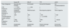
Patients with low muscle mass index, hepatic parenchyma alterations, severe tricuspid regurgitation, or advanced CKD may show altered PWD patterns in the absence of congestion, limiting the use of this technique in such cases. Table 3 summarizes the ultrasound criteria for the assessment of congestion.
- •
FoCUS: This allows for a morphological and functional assessment of the RV in different classic echocardiography planes: parasternal long axis, parasternal short axis, apical 4-chamber, and subcostal views.
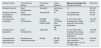
Summary of ultrasound criteria to assess congestion with LUS and VExUS techniques.
| No congestion | Mild-moderate congestion | Severe congestion | Limitations | |
|---|---|---|---|---|
| LUS | Absence of B-lines | > 3 B-lines | > 3 B-lines | B-lines in non-congestion situations: • Pneumonia • Acute Respiratory Distress Syndrome (ARDS) • Pulmonary fibrosis |
| VExUS | [4.0] Alteration in PWD patterns in non-congestion situations: • Low muscle mass • Hepatic parenchyma alterations • Severe tricuspid insufficiency • Advanced CKD | |||
| IVC | Diameter <2 cm | Diameter >2 cm | Diameter >2 cm | |
| HV | S > D | S < D | Reversed S | |
| PV | Continuous flow or pulsatility index <30% | Pulsatility index 30–50% | Pulsatility index >50% | |
| IRV | Continuous flow | S–D biphasic flow | D monophasic flow |
DP: color Doppler; LUS: lung ultrasound; IVC: inferior vena cava; VExUS: venous excess ultrasound; PV: portal vein; IRV: intrarenal veins; HV: hepatic veins.
By comparing the size of the RV with the LV and assessing septal motion, alterations in RV volume and filling pressure can be described. In these same classic planes, the systolic function of both ventricles can be evaluated relatively easily through direct visualization or using tools for estimation. A widely used and simple indirect measure to quantify RV function is measuring the Tricuspid Annular Plane Systolic Excursion (TAPSE) in M-mode. Diastolic function can also be used to evaluate volume status. FoCUS additionally allows for a rapid assessment of the presence of pericardial effusion and valvular alterations such as tricuspid regurgitation.65
The utility of PoCUS lies in the joint interpretation of the various ultrasound parameters, as they present more limitations in clinical practice when assessed in isolation. In this regard, several studies have been published demonstrating the prediction of congestive kidney injury using the different ultrasound components of VExUS, playing an especially relevant role in post-cardiac surgery patients.64 Similarly, an improvement in signs of congestion has been observed in parallel with the recovery of renal function once AKI is already established.
BiomarkersNT-ProBNPAmino-terminal pro-B-type natriuretic peptide (NT-proBNP) is the most widely used biomarker for the diagnosis and prognosis of HF. It rises due to the stress placed on cardiomyocytes in situations of increased left-sided filling pressures; however, its relationship with the severity of congestion is debatable.
The highest NT-proBNP values are observed in patients with HF due to LV systolic dysfunction, whereas right-sided dysfunction does not translate into a greater increase in the marker. Several studies show a weaker association of this marker with various clinical, radiological, and echocardiographic parameters of right-sided dysfunction.66
Other factors that can influence the increase in NT-proBNP include age and renal function, so its utility for assessing congestion in this patient group is limited.
CA-125Carbohydrate antigen 125 (CA-125) is a glycoprotein synthesized by mesothelial cells on serous surfaces and rises in response to elevated hydrostatic pressure, mechanical stress, and inflammatory stimuli. It is widely used for monitoring ovarian cancer, but it has also been observed to rise in other neoplasms and benign conditions related to volume expansion. In recent years, its use has been developed as a useful marker for identifying patients with both tissue and vascular congestion. Multiple studies associate increased CA-125 values with the presence of serous effusions, peripheral edema, increased IVC pressures, and pulmonary capillary wedge pressure.66
In contrast to NT-proBNP, CA-125 levels are not modified by renal function, age, ischemic etiology, atrial fibrillation, or LV ejection fraction. Its use for monitoring response to diuretic treatment is also relevant.67
In routine clinical practice, it is important to consider that there is a time interval between the onset of congestion and the increase in CA-125 production and release, and that it also has a long circulating half-life (7–12 days). Consequently, its utility is limited in more acute onsets with predominantly intravascular redistribution. Similarly, for monitoring the improvement of congestion, it is useful in the first weeks rather than during the first days of decompensation.68 The use of CA-125 in conjunction with other congestion parameters has proven to be an independent predictor of renal congestion measured by PWD (IRVF), which helps identify patients who would benefit from a more aggressive diuretic strategy.69
- •
Congestive nephropathy is a reversible entity defined by the triad of renal function impairment, venous congestion, and renal hypoperfusion.
- •
Outflow blood flow is compromised due to systemic congestion, with a subsequent decrease in perfusion and intratubular flow, in addition to RAAS activation, leading to tubular damage secondary to inflammatory mechanisms.
- •
PoCUS includes three strategies: lung ultrasound (LUS), which provides information on tissue congestion; the assessment of vascular congestion using the Venous Excess Ultrasound Grading System (VExUS); and the study of cardiac and valvular morphology and function through Focused Cardiac Ultrasound (FoCUS).
- •
CA-125 is positioned as a useful and potentially superior parameter to NT-proBNP for evaluating congestion, with independent predictive value for renal congestion measured by PWD (IRVF). The combined use of both biomarkers could provide complementary pathophysiological information, with CA-125 as a marker of congestion and NT-proBNP as a marker of LV functional impact.
- •
The integration of imaging techniques (PoCUS) and circulating biomarkers (CA-125 and NT-proBNP), together with clinical history and physical examination, can improve the diagnostic accuracy of congestive status, determining the predominant congestion phenotype (tissue or vascular).
One of the most frequent complications in burn patients is the development of AKI. In fact, despite improvements in burn care, AKI occurs in 50.5% of cases.70 Its importance lies in the high morbidity and mortality it entails.
Until the introduction of the RIFLE, AKIN, and KDIGO definitions, there was significant heterogeneity in the incidence of AKI in burn patients. The classification of AKI in this population using current functional criteria has shown a strong correlation between the severity of AKI and the development of unfavorable outcomes.71 Studies conducted on burn patients face an additional complication, as the patient profile varies significantly depending on the total body surface area (TBSA) burned.
In general, the incidence is estimated to be lower than in non-burn critically ill patients, primarily due to a lower average age.72
In the burn patient, two types of AKI can be distinguished based on the timing of onset.73
Early AKISome authors consider early AKI as that which develops at the time of admission, others extend it to the first 3 days, and in some cases, up to the first week. The main risk factors for its development are intravascular hypovolemia, low cardiac output, and systemic vasoconstriction during the initial resuscitation period. Therefore, the percentage of TBSA is a major risk factor. It appears that the incidence of this type of AKI has decreased in recent years due to improvements in initial management. Nevertheless, the presence of AKI upon admission can increase mortality by up to 80%.74
Late AKIThis is considered to be AKI that develops between day 2 and day 14 of admission. The primary risk factors for its development include sepsis, multi-organ failure (MOF), and the use of nephrotoxic agents.
Other risk factors that appear to influence the development of AKI are age, hypertension, diabetes mellitus, mechanical ventilation, burn mechanism (flame), inhalation injury, and prognostic scores such as the Sequential Organ Failure Assessment (SOFA).71
The diagnosis of AKI in burn patients is a factor for poor prognosis related to increased mortality. Mortality rates as high as 80% (with an average of 43%) have been described, varying according to the inclusion criteria used in different studies. Late-onset AKI has been associated with higher mortality. The development of AKI in burn patients is also linked to long-term mortality (one year post-burn), at approximately 35%.73
Furthermore, the development of AKI is associated with increased hospital stays, the need for RRT (12%), and a 2.4-fold higher risk of developing CKD, with a higher incidence in women.75
- •
There is great variability in the figures for AKI incidence, mortality, and the need for RRT due to the lack of studies with unified patient inclusion criteria.
- •
Two types of AKI are distinguished by the time of onset. Late AKI is associated with higher mortality.
- •
AKI in the burn patient is associated with higher medium-term mortality, the need for RRT, and the development of CKD.
AKI is a frequent complication in patients with liver cirrhosis (LC) admitted for acute decompensation, with an incidence of up to 50%.76 Its importance stems from the negative impact on patient prognosis.
Definitions of AKI in LC have been adapted by the International Club of Ascites (ICA) based on KDIGO definitions, adding the concepts of progression (to more severe stages), regression (to less severe stages), non-response (no regression), partial response (regression of stage with creatinine ≥0.3 mg/dL above baseline), and total response (regression of stage with creatinine <0.3 mg/dL above baseline). Additionally, the following modifications to KDIGO have been included76,77:
- 1
Urine output criteria have been removed due to the typical oliguria of cirrhotic patients secondary to avid sodium retention, the influence of diuretic treatment on output, and the limitations of precise monitoring in general wards. However, since oliguria is a sensitive and early marker of AKI and can be associated with a poorer prognosis, close monitoring should be performed whenever possible. Notably, the 2024 joint document from the ICA and the Acute Disease Quality Initiative (ADQI) also considers a urine output of ≤0.5 mL/kg for a period of 6 h as an AKI criterion in cirrhosis.78
- 2
Subdivision of KDIGO Stage 1 into two: 1A if serum creatinine (SCr) is <1.5 mg/dL (the classic cutoff for AKI in LC) and 1B if SCr is ≥1.5 mg/dL. This is based on evidence that cirrhotic patients with Stage 1 AKI and SCr 1.5 mg/dL have a significantly worse prognosis.76
- 3
Renal dysfunction may be underestimated using SCr, as hepatic production is compromised, and many patients present with sarcopenia and malnutrition. Furthermore, bilirubin elevation can interfere with colorimetric assays. Despite these limitations, changes in SCr remain a valid clinical tool. In the absence of a baseline value (community-acquired AKI), the recommendation is to use a value from the previous week; if unavailable, the ICA recommends using a value from the last 3 months instead of the 75 mL/min/1.73 m² eGFR estimation suggested by KDIGO.
The most frequent causes of AKI are prerenal (constituting up to 70%), hepatorenal syndrome (HRS), and ATN. Obstructive causes are relatively rare.
HRS is currently classified as:
- 1
HRS-AKI: When AKI criteria are met in a cirrhotic patient with ascites, there is no response to diuretic withdrawal and 48 h of albumin expansion, and in the absence of shock, nephrotoxins, proteinuria >500 mg/day, hematuria >50 RBCs/hpf, or ultrasound abnormalities
- 2
HRS-non-AKI: When AKI criteria are not met; this includes HRS-CKD (chronic kidney disease) and HRS-AKD (acute kidney disease, renal dysfunction <3 months).
Diagnostically, since prerenal causes mostly resolve with volume expansion and HRS has specific treatment unlike ATN, the priority is distinguishing these entities. FeNa is difficult to interpret; while it may be artificially elevated by diuretics, most patients, especially those with ascites, retain sodium avidly, showing FeNa <1% even with ATN. A stricter threshold (<0.2−0.5%) may increase specificity for HRS.79
Glomerular etiology should be excluded by the absence of hematuria and proteinuria. However, the cutoff for non-physiological proteinuria in LC is not well established, and serum protein levels are often low due to liver failure or malnutrition. If in doubt, a renal biopsy is required, though this is difficult due to coagulopathy. This has driven research into biomarkers; urinary NGAL can clearly differentiate HRS (normal values) from ATN (elevated values) with an AUC of 0.87 when measured on the second day of albumin expansion. Furthermore, urinary NGAL levels are associated with persistent AKI and higher 28-day mortality.80
The ICA published a sequential practical algorithm for the differential diagnosis and management of AKI in LC.77 First, precipitating factors such as volume depletion (diuretics, hemorrhage, excessive paracentesis), nephrotoxins (especially NSAIDs), beta-blockers, and infections must be corrected. For Stage 1B or higher (de novo or progressed from 1A), diuretics should be discontinued and plasma expanded with albumin (1 g/kg for 2 days). If there is no response and HRS criteria are met, vasoconstrictors (e.g., terlipressin) with albumin should be initiated. ATN or other causes should be treated individually.
By the third day, 14% of AKI cases progress, 37% stabilize, and 49% resolve.80 By hospital discharge, 67% of cases resolve, especially those due to hypovolemia or HRS-AKI.80 Nevertheless, the medium-term prognosis is poor, with a 3-month mortality rate of 60%. RRT has a clear negative impact, with median transplant-free survival reported at 15 days for HRS and 14 days for ATN.81
- •
The definition of AKI in LC differs from KDIGO: it includes evolutionary concepts, removes oliguria as a primary criterion, and subdivides Stage 1 based on absolute SCr values.
- •
The fundamental challenge is establishing a differential diagnosis to initiate correct therapy. Difficulties with standard tools have driven the use of biomarkers like NGAL to distinguish ATN from HRS.
- •
Identifying and correcting precipitating factors and applying structured early treatment can help reverse renal dysfunction.
- •
The medium-term prognosis is poor, with high associated mortality that increases when RRT is required.
The development of AKI in the context of acute HF decompensation corresponds to the definition of Type 1 Cardiorenal Syndrome.
According to various series, the incidence of AKI in this context ranges between 25% and 30%, reaching as high as 60% in patients with CKD.82
The onset of AKI has a marked prognostic value, increasing the morbidity and mortality of these patients.
Pathogenesis involves several mechanisms:
- •
Decreased cardiac output along with venous congestion, which increases renal venous and interstitial pressure, leading to a reduction in intrarenal flow.
- •
Activation of the sympathetic nervous system and the RAAS, resulting in vasoconstriction and worsening congestion.
- •
A pro-inflammatory state that induces oxidative stress affecting both renal and myocardial cells.66
Diagnosis continues to be performed through the classic parameters of decreased urine output and increased creatinine. The latter has limitations, as congestive states can decrease its sensitivity through dilution. Some studies suggest cystatin C as more sensitive and having prognostic implications. The use of renal biomarkers for early diagnosis and assessment of structural damage has been investigated, but they are not currently included in general clinical practice.83
Congestion markers NT-proBNP and CA-125 are included in the diagnostic workup (see section 4.2.2).
Management is based on adequate decongestive therapy supported by the use of potent diuretics such as loop diuretics, with the association of other diuretics if an optimal response is not achieved at high doses. Most protocols associate thiazides as a first step and aldosterone antagonists as a second step. Less common is the association of tolvaptan or the use of hypertonic saline boluses in refractory situations, as described in some protocols (see section 3.2).
If targets are not achieved, ultrafiltration techniques—with or without dialysis—would be indicated depending on the stage of AKI and associated metabolic disturbances.
Ultrasound monitoring and the assessment of congestion markers are frequently used to guide decongestive therapy. Throughout this process, the standard trend is to discontinue RAAS blockade, but some experts now suggest the possibility of maintaining treatment in cases of mild AKI, provided the patient is closely monitored.84
An increase in creatinine is relatively common during decongestive therapy. Studies report that up to 50% of patients may experience creatinine increases during therapy; expert recommendations indicate that increases < 0.5 mg/dL might signify hemoconcentration rather than true AKI. An increasing number of studies demonstrate no worsening of prognosis in patients with variable creatinine increases if adequate decongestion targets are met.85 In this regard, a decrease in filtration rate accompanied by data of hemoconcentration (increased hemoglobin and albumin) as well as a decrease in pro-BNP levels are not associated with a poorer prognosis.9,86,87 In any case, it is always appropriate in this situation to rule out other causes of AKI that may overlap in hospitalized patients.
- •
High incidence of AKI in the context of HF (CRS Type 1), around 30%.
- •
Negative effect of AKI on the morbidity and mortality of these patients.
- •
Decongestive therapy is the cornerstone of treatment, regardless of its effect on creatinine, provided the increase does not exceed 0.5 mg/dL.
- •
The use of ultrasound and congestion markers is recommended for the diagnosis and monitoring of depleting therapy.
Sepsis is the most frequent underlying cause of AKI in intensive care units, accounting for 45%–70% of cases.88 Furthermore, the association between sepsis and AKI confers a poor prognosis, with higher morbidity and mortality, longer hospital stays, and long-term sequelae such as CKD. Since both conditions are relatively common and the pathophysiology of AKI in sepsis is complex, definitions have been proposed to further study its pathogenesis, epidemiology, and progression. Thus, the **Acute Disease Quality Initiative (ADQI)**89 group has proposed the following definitions: (1) Sepsis-associated AKI (SA-AKI) when sepsis (defined by Sepsis-3)90 and AKI (defined by KDIGO, see section 1.1) coexist and appear within 7 days of the sepsis diagnosis; (2) Sepsis-induced AKI when sepsis is the primary cause of renal injury. Additionally, a distinction is made between early AKI, occurring within the first 48 h following the sepsis diagnosis, and late AKI, occurring between 48 h and 7 days.
The epidemiology of SA-AKI is essentially unknown due to the variety of definitions used across studies for both sepsis and AKI. Moreover, most studies do not differentiate between sepsis and septic shock and fail to define the temporal relationship between the two conditions.88 Identified risk factors for AKI in sepsis include septic shock, mechanical ventilation, vasopressor use, Gram-negative bacteremia, the use of RAAS inhibitors, and pre-existing conditions such as hypertension, CKD, chronic liver disease, and smoking.88
The pathophysiology of SA-AKI is complex for several reasons. First, the expression of sepsis itself depends on individual susceptibility, which is at times determined by genetic and epigenetic factors. Second, organ dysfunction associated with sepsis, including renal dysfunction, is conditioned by various physiological aspects of organs and tissues. These include micro- and macrocirculatory alterations, inflammation, immunomodulation, complement activation, RAAS dysfunction, and metabolic changes such as metabolic reprogramming and mitochondrial dysfunction. All of these factors affect the kidney in uneven and variable ways, shaping what is known as sepsis-induced AKI. Finally, other common situations in the clinical context of sepsis can affect renal function, including nephrotoxins, abdominal compartment syndrome, and congestion. These, along with the aforementioned factors, constitute the more generic term sepsis-associated AKI. In any case, sepsis-induced AKI is a distinct disorder from classic ATN, differing in its pathophysiology, circulatory dysfunction, and progression.
Given that the fundamental treatment for sepsis is the control of the source of infection, the management of SA-AKI or sepsis-induced AKI in the critically ill patient is similar to that of general AKI in the intensive care setting. Fluid administration must be restricted to the restoration of volemia to preserve tissue perfusion and should be guided by both static and dynamic parameters to avoid congestion. Hemodynamic resuscitation should be performed with crystalloid solutions, using either normal saline or balanced solutions, always based on the monitoring of electrolytic parameters. The use of albumin or bicarbonate may be beneficial in certain situations, although their recommendation requires results from ongoing clinical trials. Conversely, the use of synthetic colloids is contraindicated.91 The first-choice vasopressor in sepsis is norepinephrine, which plays a crucial role as a volume-sparing agent. The use of diuretics is restricted to the treatment of congestion.
Hemodynamic monitoring and the calculation of daily fluid balances are essential to avoid under- or over-hydration. Regarding the volume of fluids to be administered, a restrictive volume strategy has been evaluated in recent years due to the deleterious effects of secondary congestion. Various clinical trials have shown that this strategy is safe in sepsis compared to standard care. However, it does not provide significant benefits in terms of mortality, quality of life, or other relevant variables, such as the incidence of AKI, the need for RRT, or its duration.92 At the end of the observation period (90 days), the cumulative balance difference between the two strategies is approximately 0.7 l (median 1,645 mL in the restrictive group vs. 2,420 mL in the liberal group). Thus, the fluid management strategy in sepsis should follow the conceptual framework divided into different phases based on the evolutionary stage of sepsis: Resuscitation, Optimization, Stabilization, and De-escalation (ROSD), with individualization being important in all cases.89
The application of renal injury biomarkers for early diagnosis of AKI, its subclassification, or to predict the need for RRT or patient outcomes faces the same limitations as AKI from other causes.89
Regarding extracorporeal techniques, the indications and modalities for RRT are similar to those for any other type of AKI. Extracorporeal therapies aimed at modulating inflammation and the immune response are numerous (hemoadsorption, high cut-off) and can be coupled with RRT. However, recommendations for their use are very limited as there is currently no solid evidence of the benefit of these techniques on hard outcomes.
Finally, despite multiple studies, no specific treatment has been found to modify the course of AKI or the patient’s outcome in sepsis. A significant number of products have been tested, most notably dexamethasone, alkaline phosphatase, angiotensin II, levosimendan, and mesenchymal stem cell therapy.89
- •
Sepsis is the leading cause of AKI in critically ill patients.
- •
For better patient phenotyping, it is recommended to differentiate between sepsis-associated AKI and sepsis-induced AKI, as well as between early and late AKI.
- •
The pathophysiology of sepsis-induced AKI is complex and distinct from acute tubular necrosis.
- •
The treatment of sepsis-associated AKI is based on the control of the infection source along with hemodynamic management adapted to its different evolutionary stages.
The incidence of AKI in patients undergoing major surgery varies between 12% and 25% in non-cardiac surgery93 and up to 35% in cardiac surgery.94
AKI in postoperative patients results from multiple causes, including hemodynamic alterations that compromise renal perfusion (both ischemia and congestion), the use of nephrotoxins, and sepsis. In most cases, it is classified as ATN. Regarding cardiac surgery with cardiopulmonary bypass, this belongs to a special group where four major factors must be considered: the mismatch between oxygen supply and demand, the activation of the systemic inflammatory response, hemolysis, and the production of microemboli.95
The measures proposed by the 2012 KDIGO guidelines for its prevention include close monitoring of creatinine and urine output, optimization of hemodynamic parameters and volemia—for which they recommend considering dynamic or functional monitoring based on algorithms—glycemic control, and the suspension of RAAS inhibitors (ACEIs, ARBs) and nephrotoxic treatments.96 A study published by Meersch et al. in 2017 demonstrated that applying these measures in cardiac surgery patients at risk of AKI significantly reduced its occurrence.97 In the trial, patients at risk were selected through the determination of TIMP-2 and IGFBP7 biomarkers (Nephrocheck®), demonstrating that it is possible to modify the course of postoperative AKI in at-risk patients if early intervention is carried out using KDIGO recommended measures.
Furthermore, the blood pressure target in these patients is controversial and depends on their comorbidities. Several studies have shown that strict perioperative hemodynamic control is effective in preventing AKI, whether applied before, during, or early after the intervention.98 Notably, the benefit of hemodynamic control in AKI prevention is primarily obtained in high-risk surgical patients (based on clinical comorbidity scales).
Regarding the need for RRT, the initiation criteria do not differ from the classic criteria, although special attention should be paid to positive fluid balance in postoperative cardiac surgery patients.
- •
In patients at risk of AKI, close monitoring of creatinine and urine output, optimization of hemodynamic parameters and volemia, and the suspension of RAAS inhibitors (ACEIs, ARBs) and nephrotoxic treatments are recommended.
- •
Strict perioperative blood pressure control based on comorbidities is recommended for patients at risk of AKI.
- •
RRT requirements follow classic criteria, with special attention to positive fluid balance in the postoperative period of cardiac surgery.
AKI occurring during pregnancy constitutes a major public health problem due to its impact on maternal-perinatal morbidity and mortality. It is associated with a higher risk of obstetric complications: uterine hemorrhage, HELLP syndrome, abruptio placentae, and cesarean delivery. In the long term, it increases both cardiovascular risk and the risk of developing CKD. It may also increase maternal mortality. Regarding fetal prognosis, it has been associated with a higher risk of prematurity, low weight for gestational age, increased risk of neonatal ICU admission, and higher mortality.99,100
Globally, approximately 1% of AKI cases are related to pregnancy, with this frequency being higher in developing countries than in developed ones: 3.1% vs. 0.3%. In developing countries, uterine hemorrhage, septic abortions, and puerperal sepsis are the most frequent causes of pregnancy-associated AKI. In contrast, in developed countries, hypertensive disorders of pregnancy are responsible for the majority of cases.99
The definition of AKI in pregnancy is controversial, largely due to the absence of standardized diagnostic criteria. For its definition, different serum creatinine cut-off points or classification systems such as RIFLE or AKIN have been used, based on percentage increases in SCr relative to baseline. Hall and Conti-Ramsden proposed using the Kidney Disease: Improving Global Outcomes (KDIGO) definition, considering an absolute increase in SCr ≥0.3 mg/dL, within a 48-h period as the diagnosis of AKI in pregnancy.101 In this document, by consensus of all authors and based on the definition of acute kidney injury provided by the International Society for the Study of Hypertension in Pregnancy (ISSHP) as a diagnostic criterion for preeclampsia, we define AKI in pregnancy when SCr >1 mg/dL in a pregnant woman with previously normal renal function.102
A key aspect complicating the definition of AKI in pregnant women is the physiological changes that occur during pregnancy: increased cardiac output, renal plasma flow, and intrarenal vasodilation. These changes lead to glomerular hyperfiltration and a 40%–60% increase in the glomerular filtration rate (GFR). This results in a physiological reduction of creatinine, such that in most pregnant women, it remains below 0.8 mg/dL, with levels of 0.4–0.6 mg/dL being frequently found.99,103 Accordingly, formulas estimating GFR from creatinine, as well as AKI diagnostic scales (RIFLE, AKIN, KDIGO), are not validated in pregnancy. Despite all limitations, SCr remains the most cost-effective marker for diagnosing AKI. Furthermore, the lack of routine renal function monitoring during pregnancy and the frequent unavailability of a pre-pregnancy baseline creatinine complicate and delay diagnosis. Renal ultrasound can assist in identifying the etiology of AKI, while renal biopsy is rarely indicated in the pregnant woman with AKI.104
As in the general population, AKI in pregnant women is classified into three groups based on the underlying cause: prerenal, renal, and postrenal.104 Any cause of AKI occurring in the general population can happen in a pregnant woman, added to specific pregnancy-associated causes (Table 4).
Main causes of AKI in pregnancy and the puerperium.
| Prerenal: |
|---|
| a) Hyperemesis gravidarum |
| b) Obstetric hemorrhages: abruptio placentae, placenta previa, accreta, abortions, uterine atony, or rupture |
| c)Sepsis: septic abortions, puerperal sepsis, AIN, chorioamnionitis, retention of placental remains |
| d) Congestive HF |
| Renal or parenchymal: |
| a) Acute Tubular Necrosis (ATN): |
| • Severe uterine hemorrhages: uterine rupture or abruptio placentae |
| • Sepsis |
| • Drugs |
| b) Preeclampsia/HELLP syndrome |
| c) TMA: HUS, TTP, and DIC |
| d) Acute fatty liver of pregnancy |
| e) Cortical necrosis (represents approximately 5−10% of all causes of AKI in pregnancy). It may occur in the context of sepsis, abruptio placentae, uterine hemorrhages, intrauterine fetal death, TMA, DIC, and amniotic fluid embolism |
| f) Acute interstitial nephritis (AIN)/immunoallergic nephritis |
| g) Pyelonephritis |
| h) Glomerular diseases: lupus, antiphospholipid syndrome |
| i) Pulmonary embolism |
| j) Amniotic fluid embolism |
| Postrenal: |
| a) Hydronephrosis secondary to compression of the ureter or bladder by the gravid uterus |
| b) Nephrolithiasis |
| Iatrogenic or spontaneous involvement of the ureter or bladder during delivery/cesarean section |
DIC: disseminated intravascular coagulation; HF: heart failure; AKI: acute kidney injury; TMA: thrombotic microangiopathy; AIN: acute interstitial nephritis; TTP: thrombotic thrombocytopenic purpura; HUS: hemolytic uremic syndrome.
The timing of AKI onset relative to the gestational week can help establish its etiology.105 In the first trimester, the most frequent causes are hyperemesis gravidarum, septic abortion, acute pyelonephritis (APN), uterine hemorrhage, and various forms of glomerulonephritis. The latter could debut or flare throughout the pregnancy, as is the case with urinary tract infections. Undoubtedly, most causes of AKI occur in the third trimester: uterine hemorrhages associated with abruptio placentae, uterine rupture, urinary obstruction, preeclampsia (which can develop from the 20th gestational week onwards), HELLP syndrome, acute fatty liver of pregnancy, and thrombotic microangiopathies (TMA)—with an earlier onset of thrombotic thrombocytopenic purpura (TTP), which may appear from the second trimester, and a later onset of atypical hemolytic uremic syndrome (aHUS), more frequent in the peripartum and postpartum periods (Table 5). Finally, in the puerperium, AKI associated with uterine hemorrhage (atony, uterine perforation), puerperal sepsis, NSAID nephrotoxicity, and the aforementioned aHUS stands out.105,106
Differential diagnosis of TMA in pregnancy.
| Severe PE | HELLP | TTP | aHUS | |
|---|---|---|---|---|
| Time of diagnosis | Generally 3rd trimester | 3rd trimester | 2nd and 3rd trimesters | 3rd trimester/postpartum |
| HTN | + + + 100% | + / + + 80% | ± | + + |
| Proteinuria | Very frequent | + / + + | + | + / + + |
| Common symptoms | Headache, epigastralgia | Abdominal pain | Neurological symptoms | Renal, digestive |
| Hemolytic A. | + /0 | + | + + | + |
| LDH U/l | <600 | >600 | >1.000 | >1.000 |
| Thrombocytopenia | + | + + | + + + (< 20.000) | + + |
| Hepatic cytolysis | + | + + + | ± | ± |
| AKI | ± Mild | + Mild | ± Mild | + + Severe |
| Postpartum recovery | 48−72 h | < 1 week | No | No |
| Treatment | Symptomatic Delivery | Symptomatic Delivery | Plasma exchange Corticoids | Eculizumab Ravulizumab |
HELLP: Hemolysis, Elevated liver enzymes, Low platelet; HTN: hypertension; AKI: acute kidney injury; LDH: lactate dehydrogenase; TMA: thrombotic microangiopathy; PE: preeclampsia; TTP: thrombotic thrombocytopenic purpura; aHUS: atypical hemolytic uremic syndrome.
Management of pregnancy-associated AKI includes:
- 1
General measures: Fluid therapy for volume depletion, antibiotics for sepsis, transfusion for severe hemorrhage, diuretics for HF, blood pressure control, correction of electrolyte and acid-base imbalances, and dialysis when conservative management is insufficient.
- a
Preeclampsia with severity criteria, HELLP syndrome, acute fatty liver of pregnancy: Supportive measures as a bridge to the only effective treatment: induction of labor
- b
Hyperemesis gravidarum: Rehydration
- c
TTP: Plasmapheresis, steroids, rituximab, caplacizumab
- d
aHUS: Eculizumab/ravulizumab
- e
Obstructive uropathy: Urinary tract diversion
- f
Abruptio placentae: Bleeding control and urgent delivery.
- g
Glomerulonephritis: Steroids and immunosuppressants compatible with pregnancy
- a
- •
Pregnancy-associated AKI increases maternal-perinatal morbidity and mortality.
- •
It is underdiagnosed.
- •
Renal function should be monitored more closely during pregnancy: quarterly and whenever the pregnant woman presents any complication that could be associated with AKI.
- •
Early diagnosis is required, fundamentally in cases where failure to initiate rapid and effective treatment could lead to advanced CKD.
- •
Management by a multidisciplinary team is necessary, in close collaboration with the Obstetrics and Anesthesiology services.
The substances that can cause AKI through toxicity are numerous, with highly varied chemical and biological characteristics. However, their nature varies depending on the setting of exposure. While in the hospital setting it is almost exclusively related to drug use, in the outpatient setting, non-pharmacological agents such as drugs of abuse, medicinal herbs, heavy metals, alcohols, or certain pesticides must also be included.
The incidence varies according to the series and diagnostic criteria. The most studied form is pharmacological toxicity, which causes approximately 16%–29% of hospital-acquired AKI.107 Determining the outpatient incidence is more complex, with estimated figures ranging from 59% to 72%, increasing in elderly and polymedicated patients.108
Potentially nephrotoxic substances or their metabolites are eliminated by the kidneys through glomerular filtration, tubular secretion, or a combination of both, exposing tubular cells to risk. Several mechanisms are involved in this damage109:
- 1
Direct tubular toxicity due to intracellular accumulation of substances eliminated by glomerular filtration with tubular reabsorption.
- 2
Toxicity from components eliminated via tubular secretion from the basolateral membrane, causing metabolic interference leading to necrosis and apoptosis.
Intratubular crystal deposition resulting in obstruction and cellular injury.
Intratubular cast formation.
Alterations in intrarenal hemodynamics.
Immunoallergic reactions such as AIN.
Rhabdomyolysis.
Thrombotic microangiopathy (TMA).
Regardless of the toxin's characteristics and particularly in the case of drugs, several patient-dependent factors can favor the onset of toxicity. Some are potentially modifiable, such as hydration status, metabolic and acid-base imbalances, pre-existing CKD, cardiorenal syndrome, nephrotic syndrome, or cirrhosis. Others are non-modifiable, most notably age, female sex, and pharmacogenetic alterations that affect the metabolism and renal handling of drugs.
In addition to the non-pharmacological substances previously mentioned, the drug groups most frequently implicated in AKI are antibiotics, NSAIDs, antivirals, RAAS blockers, and both classic and next-generation chemotherapeutic molecules.
Immunoallergic nephritis (AIN), however, can be triggered by any type of drug.110 In this section, new antineoplastic immune checkpoint inhibitors deserve mention, as one of their most significant side effects is renal involvement—predominantly expressed as immune-mediated nephritis—representing a new challenge given the widespread use of these treatments.111
The diagnostic approach utilizes standard parameters such as SCr elevation, despite its limitations for early diagnosis, as well as urine assessment. The latter may reveal tubular-type proteinuria with a predominance of low-molecular-weight proteins and sediment alterations such as hematuria and/or leukocyturia, which are more frequent in AIN (especially sterile leukocyturia, although in various series this does not exceed 50% of cases). Other urinary findings may include crystals, though they are more commonly observed in renal biopsies. Biopsies are necessary primarily to detect immunological phenomena that require specific treatment. There is no evidence yet for the routine use of early kidney injury biomarkers, although the FDA has approved the use of KIM-1 as a marker of nephrotoxicity for certain drugs.
Treatment must include withdrawal of the toxin. General measures include adequate hemodynamic support and fluid balance aimed at ensuring correct urinary flow (essential in crystal-induced injury), and correction of electrolyte and acid-base imbalances. More specific measures include the use of antidotes or chelators in the case of heavy metals. Steroid use is relevant in AIN, where an early start is fundamental to reducing established lesions. Finally, extracorporeal clearance techniques may be used, either as support for AKI with standard indications or to eliminate dialyzable substances, as in alcohol poisoning.111
Prevention is based on:
- 1
Appropriate indication of potentially nephrotoxic drugs, avoiding combinations that may potentiate harmful effects.
- 2
Adjusting drug doses according to the GFR.
- 3
Maintaining or restoring adequate hydration status with proper urinary flow.
- 4
Correcting metabolic and acid-base imbalances and appropriate monitoring of renal function.
Lastly, it should be noted that toxic AKI can lead to a poor renal prognosis, as significant lesions and delayed diagnoses increase the risk of CKD.
AKI and hemorrhagic shockHemorrhagic shock is a pathophysiological state caused by the rapid and significant loss of intravascular volume, which leads sequentially to hemodynamic instability, inadequate tissue perfusion, microcirculation alterations, cellular hypoxia, cellular damage, systemic inflammation, and multi-organ dysfunction, potentially resulting in patient death. The complexity of the condition depends on the volume of blood lost and the speed of its onset.
The prevalence of AKI in the context of hemorrhagic shock is related to the severity of the hemorrhage. Typically, the kidneys are affected when the loss of circulating volume exceeds 20%–40%. In more than 90% of cases, renal dysfunction appears within the first 5 days after the event, being most frequent within the first 48–72 h. If we use blood transfusions as a marker of bleeding, AKI appears in 28.3% of patients requiring at least one unit of packed red blood cells and reaches 42.5% in patients presenting with hemorrhagic shock. The development of AKI has been independently associated with an increased risk of mortality in these patients within the ICU.112,113
Various studies have identified age, high comorbidity burden, severe hypotension, tachycardia, time elapsed before reaching the hospital (in trauma cases), elevated serum lactate and troponin levels, thrombocytopenia, and a hemodynamic state of shock as predictive factors for AKI in this context. Blood lactate, even in the absence of hypotension, acts as a predictor by being a marker of tissue hypoperfusion. In trauma patients, the hemodynamic situation is compounded by potential severe rhabdomyolysis, where the creatine kinase level acts as an independent factor for the development of AKI.112,113
Hemodynamic instability is a risk factor for the development of AKI. Pathophysiological mechanisms include a drop in renal perfusion pressure and microcirculation impairment. Although restoring mean arterial pressure (MAP) may mitigate the risk of renal injury to some extent, the evidence remains contradictory. Classically, it is stated that renal injury responds to a decrease in renal blood flow and intense renal vasoconstriction, causing severe tissue ischemia with the release of pro-inflammatory CKs and free radicals.114 However, recent research contradicts the notion that renal injury is solely caused by vasoconstriction and subsequent tubular necrosis, postulating instead that intrarenal vasodilation exists alongside microcirculatory changes produced by vasoregulatory mechanisms that determine a vasodilatory effect on precapillary arterioles. Experimental studies show that in hypovolemic shock, the drop in shear stress causes alterations in endothelial NOS functionality, leading to increased nitric oxide availability that alters the tubuloglomerular feedback mechanism and conditions the loss of renal autoregulation after renal ischemia.115
Furthermore, hypovolemia in circulatory shock leads to activation of the sympathetic nervous system, which in turn can result in acute kidney injury. Decreased cortical perfusion during hypovolemia also leads to renin release by the juxtaglomerular apparatus, stimulating aldosterone production, which favors sodium and water retention in the renal tubule in an attempt to maintain volume.112,113
Concurrently, hemorrhagic shock initiates a vicious cycle of hypothermia, acidosis, and coagulopathy—also known as the lethal triad—which has a damaging effect on renal cells. Metabolic acidosis, resulting from cellular anaerobic metabolism secondary to tissue hypoxia, facilitates tubular injury; hypothermia leads to a decrease in glomerular filtration; and hypercoagulability, initiated in early stages to control hemorrhage, can ultimately cause glomerular thrombosis.116,117
It is crucial to rapidly identify the source of hemorrhage, take appropriate clinical decisions to control it, and initiate patient resuscitation as soon as possible. To date, research has focused on the initial resuscitation period, based on the administration of blood products and intravenous crystalloids before and simultaneously with hemorrhage control or hemodynamic stabilization. Generally, a MAP target of around 65 mmHg is recommended in the initial management of shock. In patients with hemorrhagic shock without severe brain injury, a lower MAP target is recommended. This is known as permissive hypotension, which, by decreasing intravascular pressure, minimizes active blood loss from unrepaired lesions. Although an increasing number of trials conclude that a more conservative strategy—administering minimal fluid amounts—is more effective, controversies regarding overall therapeutic management persist. On the other hand, the management of the patient once initial stabilization is achieved (post-hemostatic resuscitation) remains largely unaddressed.116,118,119
Regarding fluid therapy, rapid intravenous fluid administration is recommended until blood pressure enters the desired range. The recommendations for volume limitation and monitoring described above must be taken into account.
Regarding vasoactive drugs, the most tested therapeutic options to improve renal perfusion consist of catecholamines, vasopressin, and angiotensin II, all as volume-sparing agents. Although catecholamines are the most studied, they are associated with adverse events at higher doses, including AKI. Vasopressin and angiotensin II could be options by improving intraglomerular hemodynamics through differential vasoconstriction of efferent and afferent arterioles within the same nephron. Terlipressin may contribute to restoring hemodynamic stability and has anti-inflammatory effects. Experimental studies in rats have shown that terlipressin and vasopressin could be viable therapies for hemorrhagic shock-induced AKI, likely attenuating said injury by modulating the inflammatory response.110,120
In more initial phases are studies on the potential beneficial effects of inhibiting the sympathetic system on renal structure and function in response to ischemia, as well as the use of endothelial nitric oxide synthase (NOS) inhibitors, which have shown improvements in renal autoregulation efficiency independently of their effects on renal plasma flow.115
- •
The onset of AKI in the context of hemorrhagic shock is early, frequent, and related to the severity of the hemorrhage.
- •
The pathophysiology is multifactorial and conditioned by the severe decrease in renal perfusion, microcirculation alterations, renal autoregulation impairment, sympathetic system activation, direct tubular injury due to tissue hypoxia, and intraglomerular thrombosis.
- •
Currently, evidence regarding the therapeutic approach is limited, with numerous controversies existing both in the initial stabilization phase and once stabilization has been achieved.
- •
The current trend regarding intravenous fluid therapy is restrictive and combined with vasopressors, as this translates into better overall and renal outcomes.
The incidence of AKI in the postoperative period of solid organ transplantation is high and significantly influences morbidity, mortality, and the development of CKD in these patients, especially if they require RRT.
In lung transplantation, figures range from 39% to 80%, with 100% mortality at 2 years if dialysis was required. In heart transplantation, the incidence can reach up to 75% of cases, and in liver transplantation, with more disparate diagnostic criteria, it ranges between 12% and 80%.121
Several common and specific factors may contribute to these incidence rates.121,122
Common factors include:
- 1
The presence of pre-existing CKD, which is sometimes underestimated in these patients, for whom creatinine and the formulas estimating GFR based on it are less accurate.
- 2
Significant hemodynamic disturbances occurring during these surgeries along with postoperative volume and hemodynamic management.
Specifically, the following stand out:
- 1
In lung and heart transplantation: The need for vasopressor support, prolonged cardiopulmonary bypass time, the use of extracorporeal membrane oxygenation (ECMO), and the use of intra-aortic balloon pumps or ventricular assist devices.122
- 2
In liver transplantation: A high Model for End-Stage Liver Disease (MELD) score, presence of decompensated cirrhosis prior to the intervention, significant graft ischemia-reperfusion injury, early graft dysfunction, and donors after circulatory death. More recently, the influence of reperfusion syndrome has been described, which entails circulatory collapse with hypotension, increased pulmonary pressures, and decreased cardiac output—attributed to the acidosis, hyperkalemia, and hypothermia occurring upon portal vein unclamping. Additionally, the release of inflammatory cytokines may contribute to the damage.123
Diagnosis based on KDIGO criteria using SCr presents some difficulties, as high volume intake and transfusions can cause hemodilution and delay detection; therefore, monitoring urine output can be helpful.
The use of biomarkers is more extensively studied in cardiac surgery, especially NGAL and lately the cell cycle arrest marker TIMP2*IGFBP7, with an intervention study for AKI prevention. Some studies with NGAL in liver transplantation suggest it may be a predictor of AKI, but its use is not yet widespread.122
Treatment involves general aspects of AKI management, such as strict hemodynamic and volume control, along with specific elements like appropriate monitoring of CNI levels.
In general, the need for RRT ranges between 5% and 15% of patients with AKI, depending on the series and type of transplant, with continuous techniques being used most frequently.122
Prevention begins with a thorough understanding of the pre-transplant renal status, regarding both the accurate determination of GFR and the detection of pre-existing structural renal damage. Adequate hemodynamic support during surgery, as well as proper management of both immunosuppressive and potentially nephrotoxic medication, is especially relevant, as these patients require varied antibiotic therapies and occasionally contrast-enhanced examinations, as occurs in heart transplantation. In liver transplantation, the surgical technique with vena cava preservation reduces the incidence of reperfusion syndrome, which impacts the risk of AKI.
- •
AKI in solid organ transplantation is frequent and carries significant implications for morbidity and mortality.
- •
Prevention is difficult given the multiple and diverse factors involved.
- •
The need for renal replacement therapy (RRT) is common.
- •
AKI is involved in the progression to CKD in these patients.
The development of an AKI episode, beyond its immediate negative impact during hospitalization, also results in adverse medium- and long-term outcomes: persistent decrease in GFR over time, recurrence of AKI episodes, development or progression of CKD and end-stage renal disease (ESRD), increased cardiovascular risk, higher hospital readmission rates, and long-term mortality.
In the 2017 ADQI consensus document and the 2020 KDIGO review, the term Acute Kidney Disease (AKD) was proposed to define a clinical situation of impaired renal function that does not meet the criteria for AKI or CKD, yet is related to adverse medium- and long-term outcomes.124–126
Currently, a new conceptual framework is proposed for the spectrum of renal disease over time, conceiving AKI, AKD, and CKD as a continuum (sharing risk factors and pathophysiological mechanisms). Criteria for AKI and CKD remain those defined by existing KDIGO guidelines, although it is important to note that these do not yet include criteria for defining AKI resolution. The proposed criteria for AKD (occurring between 7 days and 3 months) include: SCr increase >50%, eGFR <60 mL/min, a drop in eGFR ≥35% from baseline (using CKD-EPI 2009 for SCr or CKD-EPI 2012 for cystatin C), or the presence of structural damage markers (mainly albuminuria or hematuria). By definition, AKD precedes CKD but can also overlap with pre-existing CKD.
The ADQI group suggests grading AKD severity according to KDIGO AKI stages to define severity and provide a framework for kidney-specific outcomes within a 90-day timeline126 (Table 6).
AKD Stages.
| Stage 0 (subacute AKD) | A: Without evidence of damage |
| B: Increase in biomarkers or loss of renal functional reserve | |
| This stage can be difficult to identify without nephrological follow-up. It includes new-onset proteinuria or worsening of existing proteinuria, as well as de novo hypertension or worsening of its control. If available, it also includes loss of renal functional reserve evidenced by nuclear medicine tests | |
| C: SCr does not reach baseline values, but its increase is less than 1.5 times the baseline value | |
| Stage1 | SCr 1.5−1.9 times the baseline value |
| Stage2 | SCr 2−2.9 times the baseline value |
| Stage3 | SCr ≥ 3 times the baseline value or need for renal replacement therapy |
AKD: Acute kidney disease; SCr: serum creatinine.
This classification aims to standardize research results and facilitate the determination of incidence and prognosis.
Markers of progressionOne reason for the increase in CKD after AKI is "lack of recovery", this is a gradual decline in eGFR that does not return to baseline. Ideally, a biomarker would allow for early diagnosis to implement management strategies aimed at preventing progression.
Using SCr has limitations due to muscle mass loss, changes in distribution volume (dilution), and hyperfiltration; it is also a late marker of injury. GFR at discharge is also a poor predictor of CKD progression. Sawhney et al. demonstrated in a cohort of 14,651 patients followed for 10 years that GFR at discharge does not correlate with CKD progression, and this risk persists for up to 10 years post-AKI.127
Regarding proteinuria as a progression marker, the ASSESS-AKI study showed that higher albumin-to-creatinine ratios (uACR) 3 months after discharge were associated with a higher risk of CKD.128 Although baseline proteinuria was unknown, uACR remains an excellent discriminative tool and a modifiable factor in clinical practice.
Although most studies on progression markers focus on identifying markers that imply AKI persistence after 48−72 h, several works have been published identifying different biomarkers (NGAL, KIM-1, CCL14, TIMP-2*IGFBP7, IL-18, MCP-1, bFGF, NT-proBNP, TNRF1, or sTNFR2) associated with longitudinal adverse outcomes following an AKI episode: mortality events, cardiovascular events, and progression to CKD.9 However, despite extensive research and the development of assays for some of them, these biomarkers remain restricted to research use and have not yet been implemented in clinical practice. It remains to be demonstrated, through prospective trials, to what extent these new biomarkers can help improve short- and long-term outcomes.
Assessment of renal function at discharge in patients after an AKI episodeThe definition of renal function recovery following an AKI episode remains controversial due to the absence of standardized criteria. Various approaches have been used in the literature, ranging from the normalization of SCr to the discontinuation of RRT. It is important, due to its prognostic impact, to differentiate whether the recovery of renal function is complete or partial, as well as the time it takes for renal function to recover. To unify criteria, the authors of this document, as representatives of the Acute Kidney Injury (AKI) workgroup of the Spanish Society of Nephrology (FRASEN), propose, in agreement with other researchers,129 to consider an AKI episode recovered when SCr returns to its baseline level, and partial recovery if it does not reach this threshold. However, even if creatinine levels and GFR return to baseline values, it must not be forgotten that a significant reduction in renal functional reserve may have occurred, exposing the patient to a higher susceptibility to future AKI episodes.130
The FRASEN group recommends assessing recovery at the time of hospital discharge by determining the SCr value. It is considered that this should always be accompanied by the determination of the albumin/creatinine ratio, and the performance of CrCl should be assessed on an individual basis as an indicator of structural renal injury.
It is important to determine the timeframe of discharge relative to the AKI episode. We must not lose sight of the fact that apparent "recovery of renal function" at discharge is not always an indicator of a good renal prognosis, as SCr at that moment may be overestimating the GFR. Furthermore, there may later be a progressive loss of renal function conditioned by numerous factors that are currently difficult to predict with the tools available in clinical practice.
Post-AKI patient follow-upCurrently, there are no standardized guidelines for the follow-up of patients with AKI, nor treatment strategies to reduce the incidence of sequelae.
During the AKI episode, the main objective should be the recovery of baseline renal function in the shortest possible time to reduce the duration and severity of the injury. After discharge, it is crucial to preserve renal function and prevent further deterioration by controlling hypertension, proteinuria, diabetes mellitus, and cardiovascular diseases.131
The KDIGO 2012 guidelines recommend follow-up at 3 months post-AKI to assess the resolution of AKI or the persistence of CKD.96,132 This point is probably very late, and the renal injury is already established. In the new KDIGO 2020 recommendations, the degree of nephrological follow-up increases as the duration and severity of AKI/AKD increase.124 According to these recommendations, we must be sensitive to those factors identified as risks for progression and focus on them to establish the timing and degree of follow-up. The risk factors for the progression of renal injury to chronic renal failure identified in previous studies are shown in Table 7.
In the opinion of the FRASEN group, the planning of follow-up visits should be organized based on the severity and duration of the AKI, the need or not for RRT during admission, the degree of recovery of the renal injury based on the indicators currently available in clinical practice, the organizational possibilities of each Nephrology Service, and the relationship with Primary Care for shared follow-up. We consider a review of the patient within the first 30 days of discharge to be optimal in the most severe cases or comorbid situations; if this is not possible, we advise planning the review at 3 months.
At the discretion of the nephrologist who treated the AKI, milder cases (with an episode duration of less than 7 days and/or with complete recovery of renal function at discharge) could be referred to primary care for follow-up. In this sense, we should work toward multidisciplinary follow-up for patients who have suffered an AKI (nephrologist, primary care, nursing, pharmacy, among others). The literature demonstrates that this type of follow-up achieves better results.133
Regarding outpatient follow-up and after reviewing the literature, the FRASEN group, taking into account the absence of evidence and the lack of international consensus, recommends the following measures:
- 1
Monitor renal function and proteinuria. Incorporation of new markers of renal injury based on new evidence generated.
- 2
Evaluate the introduction of nephroprotective medication. It will be important to assess potential discontinuations of nephroprotective drugs during the episode (ACEI, ARB, antialdosteronics, SGLT2i) for their reintroduction, according to risk/benefit, in a controlled manner.
- 3
Education in nephroprotection. The patient and their environment should be informed about nephrotoxic medications that must be avoided. Recommend the temporary discontinuation of certain medications in situations of dehydration risk (gastroenteritis or fever) to prevent new AKI episodes.
- 4
Patients who continue to require dialysis at the time of hospital discharge must be monitored during the sessions. In these patients, hemodynamic status, intravascular volume, and diuresis during dialysis must be carefully controlled so as not to interfere with the possibility of renal function recovery. Higher ultrafiltration rates and more intradialytic hypotension episodes are associated with a higher risk of non-recovery.124
- •
It is crucial to stop considering AKI as a short-term reversible condition and to raise awareness about long-term complications, such as progression to CKD, increased cardiovascular events, and mortality.
- •
Further research is required to understand the pathophysiological mechanisms involved and to identify potential renal and vital prognosis biomarkers, upon which optimal standardized follow-up and prevention strategies can be established. Looking to the future, there is a need to explore therapeutic actions that, established early, prevent the progression of renal failure.
- •
Although standardized guidelines are lacking, after an episode of severe AKI (stage 2 or 3), patients should have specialized follow-up. Nephrological follow-up is crucial to evaluate renal function after the episode, perform medication reconciliation, educate patients on the prevention of nephrotoxicity, avoid the occurrence of new AKI episodes, and implement strategies to prevent progression to CKD.
- •
In general, there is very heterogeneous and suboptimal follow-up of AKI after discharge, largely due to a lack of awareness and the absence of standards for prevention and management.
AKI is the most frequent cause for initiating RRT, both in continuous modalities and intermittent hemodialysis (IHD). Continuous treatment will be used when the patient is hemodynamically unstable or neurocritical, while an intermittent modality will be used in all other cases.136,137
Emergent indications for the initiation of RRT:
- 1
Oliguria/anuria unresponsive to standard medical treatment: volume expansion, diuretics, vasoactive drugs, inotropic agents.
Acute pulmonary edema with no response to medical treatment.
Hyperkalemia >6.5 mmol/L refractory to medical treatment.
Metabolic acidosis with pH < 7.2 refractory to medical treatment.
Retention of nitrogenous waste products with secondary uremic complications.
There are other non-emergent indications for starting RRT in the critically ill patient, where it could be considered as supportive therapy:
Metabolic control in hypercatabolic situations.
- 1
Volume control: Volume overload is an independent risk factor for mortality in critically ill patients and, on occasion, is the determining cause for initiating RRT.138 In these cases, continuous ultrafiltration facilitates the management of the critical patient, avoiding volume overload resulting from the continuous administration of intravenous drugs (antibiotics, vasoactive drugs, inotropes) and blood products, while simultaneously allowing the administration of the appropriate volume of enteral/parenteral nutrition (PN) according to their needs.
Finally, there are other situations, in which AKI may or may not be present, that may require the initiation of RRT:
- 1
Intoxications: In non-critical patients, IHD is the treatment of choice.139 However, in critical patients, there are some drug intoxications, such as lithium or metformin, where continuous RRT may be an option. These drugs tend to show rebound effects after IHD, and a slower elimination rate does not pose a serious risk to the patient. Another therapeutic scheme in these cases would be a mixed treatment: performing IHD first and subsequently continuing with a continuous technique.
- 2
Lactic acidosis: allows for the simultaneous removal of lactic acid and the provision of bicarbonate. However, in the absence of AKI or associated metformin intoxication, there is little evidence that initiating RRT influences patient prognosis.136
- 3
Electrolyte disturbances, refractory to medical treatment, in hemodynamically unstable patients or those with severe brain injury.
- 4
Congestive HF: SCUF (slow continuous ultrafiltration) as the technique of choice.140
- 5
Refractory hyper- or hypothermia.
- 6
Rhabdomyolysis: for the management of AKI, electrolyte disorders, or associated volume overload.
- 7
Major burns: hypercatabolic patients with difficulties in fluid management.
- 8
Metabolic and volume management in severe traumatic brain injury
- 9
In liver failure, when accompanied by AKI or for the management of volume overload.
- •
AKI is the most frequent cause for initiating any RRT in the critically ill patient, but it is not the only one.
- •
There are non-emergent indications for starting RRT, such as better metabolic control or adequate volume control.
- •
There are other indications in which RRT is initiated in patients without AKI, notably congestive HF, lactic acidosis, and certain types of intoxications.
If there is a topic that has sparked controversy regarding the management of critically ill patients with RRT, it is the optimal timing to initiate the technique and, above all, whether this has any impact on patient prognosis. The decision is clear when any of the emergent indications for starting dialysis exist: uremia, hyperkalemia, metabolic acidosis not corrected with medical treatment, or acute pulmonary edema that does not respond to diuretics. However, in the absence of these situations, there is no evidence to establish a definitive recommendation on when to initiate treatment.141
Such is the interest aroused by this subject that in the last decade, five major clinical trials have been published in high-impact journals aimed at answering this question. The ELAIN study, a single-center study conducted in Germany, concluded that early initiation of the technique was associated with significantly higher survival at 90 days of follow-up compared to delayed initiation.142 Almost in parallel, the AKIKI study, a multicenter study conducted in France with a 60-day mortality objective, found no differences.143 Furthermore, it highlighted a significant fact: there was a high percentage of patients randomized to the delayed initiation arm who, ultimately, did not require dialysis. Subsequently, the IDEAL-ICU and STARRT-AKI trials appeared, both multicenter, which also found no differences in 90-day survival when comparing early vs. later initiation, confirming once again that a non-negligible percentage of patients in the delayed initiation arm did not end up being dialyzed (38% in IDEAL-ICU, figures similar to those found in STARRT-AKI).144,145 More recently, AKIKI 2 concluded that in critically ill patients with oliguria and urea levels higher than 220 mg/dL, even in the absence of other indications for urgent dialysis, delaying the start of RRT any further does not bring benefits and does entail potential harm.146 The results of these clinical trials are summarized in Table 8.
Clinical trials on the initiation of RRT.
| Clinical Trial | Publication Year, Country, centers, RRT Mode, Population | Inclusion Criteria | Study Arms | Study Objective |
|---|---|---|---|---|
| ELAIN | 2016, Germany, single-center, continuous RRT, predominance of surgical ICU patients | AKI Stage 2 KDIGO, NGAL >150 ng/mL severe sepsis | Early: RRT <8 h Delayed: >12 h or Stage 3 KDIGO | 90-day mortality. Differences between groups, favorable toward early initiation (p = 0.03) |
| AKIKI | 2016, France, multicenter, according to treating physician's criteria, 55% IHD as initial therapy, predominance of medical ICU patients | AKI Stage 3 KDIGO, MV and/or vasoactive support | Early: RRT <6 h Delayed: oliguria >72 h | 60-day mortality. No differences between groups (p = 0.79) |
| IDEAL-ICU | 2018, France, multicenter, according to treating physician's criteria, medical/surgical ICU, septic shock | Septic shock and RIFLE Stage F | Early: RRT <12 h Delayed: >48 h | 90-day mortality. No differences between groups (p = 0.38) |
| STARRT-AKI | 2020, Canada, multicenter, according to treating physician's criteria, medical/surgical ICU | AKI Stages 2 or 3 KDIGO | Accelerated: RRT <12 h Standard >72 h | 90-day mortality. No differences between groups (p = 0.92) |
| AKIKI 2 | 2021, France, multicenter, according to treating physician's criteria, medical/surgical ICU | AKI Stage 3 KDIGO, MV and/or vasoactive support, oliguria >72 h or urea greater than 220 mg/dL | Delayed initiation: <12 h More delayed: urea greater than 220 mg/dL or emergency indication | Recovery of renal function (days free of RRT). Delayed: 12 days. More delayed: 10 days Higher 60-day mortality in the more delayed group |
ELAIN: Early versus delayed initiation of RRT on mortality in critically ill patients with AKI AKIKI: Artificial kidney initiation in kidney injury IDEAL-ICU: Initiation of dialysis early versus delayed in the intensive care unit STARRT-AKI: Standard versus accelerated initiation of RRT in AKI AKIKI 2: Comparison of two delayed strategies for RRT initiation for severe AKI; RRT: Renal replacement therapy; ICU: Intensive care units; MV: Mechanical ventilation; E2: Stage 2 KDIGO (Kidney Disease: Improving Global Outcomes) classification of ARF; E3: Stage 3 KDIGO (Kidney Disease: Improving Global Outcomes) classification of AKI; Stage F: Failure of kidney function from the RIFLE classification; AKI: Acute kidney injury; NGAL: Neutrophil gelatinase-associated lipocalin; RIFLE: risk, injury, failure, loss end-stage kidney disease.
In 2022, a meta-analysis was published including 5,193 critically ill patients with AKI with a primary objective of 28-day mortality; it concluded that the early initiation of RRT does not provide a benefit in terms of patient survival or renal function recovery and increases the risk of adverse events associated with the technique.147
In a Cochrane review also published in 2022, the authors concluded, with a low to moderate level of evidence, that early initiation of RRT in patients with AKI does not provide any benefit in terms of survival. It may have a slight benefit in the recovery of renal function and probably reduces the mean stay in the ICU, as well as the hospital stay, but it increases the risk of adverse events.148
The important thing in this matter is to use common sense and weigh the benefits derived from an early initiation of the technique (better metabolic, nutritional, hemodynamic, and volume control) against the risk of rushing and subjecting the patient to unnecessary treatment, which is not without complications. It is necessary to individualize when indicating an RRT, using clinical and hemodynamic criteria rather than biochemical ones, and to remember that what may apparently be early for one patient may be late for another and vice versa.
- •
In the critically ill patient with AKI, in the absence of emergent indications, there is no evidence to establish a definitive recommendation on when to initiate RRT.
- •
Regarding the survival of the critically ill patient with AKI, most clinical trials and meta-analyses do not find a benefit derived from the early initiation of the technique.
- •
It is important to take into account the possible benefits derived from the early initiation of RRT, but weighing the risk of rushing and subjecting the patient to unnecessary treatment that is not without complications.
- •
When initiating RRT, it is necessary to individualize, using more clinical than biochemical criteria.
Regarding the choice of vascular access for RRT in patients with AKI, the recommendation is to use a percutaneous catheter of appropriate gauge (10F or larger) and length (17 to cm) according to the site. The preferred location is the right internal jugular vein, followed by the femoral vein (in this case, long catheters of more than 20 cm are recommended), the left jugular vein, and, as a last option, the subclavian vein.96 In any case, placement must be ultrasound-guided, and the position must be verified by chest X-ray before use. If the expected duration of RRT is more than 2 weeks, the implantation of a tunneled catheter may be considered.
What type of renal replacement therapy to use in the critically ill patient with AKIRRT is key in the management of patients with severe AKI, but focusing on the critically ill patient, what is the RRT of choice? A continuous or an intermittent technique? The reality is that there is no ideal technique for all patients. Its choice will depend on the patient's hemodynamic status, the availability of techniques in each hospital, and the physician's experience with each therapeutic modality.149
Undoubtedly, in the critically ill patient, continuous RRT has important advantages derived not only from a lower ultrafiltration rate but also from a slower and continuous removal of solutes, without large fluctuations in osmolarity. All of the above leads to better vascular refilling, with a lower risk of hypovolemia and, theoretically, better hemodynamic tolerance. However, based on the results of the only clinical trial that specifically compares intermittent dialysis with continuous techniques, both techniques would be equivalent regarding the need for vasoactive drugs.150 Nevertheless, in critically ill patients, IHD has been associated with hypotension in up to 70% of cases, a frequency clearly higher than that reported with continuous techniques, which is around 45%.151,152 In addition to the advantages in hemodynamic tolerance, other benefits of continuous RRT would be the positive effect on microcirculation due to the preferential removal of fluid from the interstitial space, with secondary respiratory improvement, and the possible immunomodulatory role it may exert on the inflammatory response in sepsis.
The KDIGO guidelines for AKI management suggest that continuous RRT may be preferable to IHD in two population groups: neurocritical patients with acute brain injury, generalized cerebral edema, and other causes of increased intracranial pressure; and in hemodynamically unstable patients.2,96 The problem is that there is no established definition of hemodynamic instability when choosing the RRT modality, so it must be individualized. Thus, as a guide, intermittent techniques would be indicated as the initial technique in hemodynamically stable patients (MAP >70 mmHg, without vasoactive drugs or with norepinephrine at doses ≤0.1 µg/kg/min) or as a continuation of continuous therapy when the patient is already hemodynamically stable but with a persistent need for RRT.
Regarding the recovery of renal function or patient survival with AKI, the superiority of one technique over the other has not been demonstrated.153 In 2022, a secondary analysis of the AKIKI and IDEAL-ICU clinical trials was published—two studies comparing two RRT initiation strategies in patients with severe AKI: early vs. late initiation. The objective of this sub-analysis was to see if the RRT modality (continuous vs. IHD) influenced survival. No difference in survival was demonstrated between the two techniques.154 A year later, a secondary analysis of the Standard versus Accelerated Renal Replacement Therapy in Acute Kidney Injury (STARRT-AKI) clinical trial was published, comparing the prognosis of critically ill patients with AKI who initiated RRT with a continuous technique vs. IHD. In this sub-analysis, it was concluded that initiation with a continuous technique, compared with IHD, is associated with a significant reduction in the composite outcome of death and dialysis dependence at 90 days.149 However, although the results of this study seem to give an advantage to continuous techniques, being a sub-analysis, they must be handled with caution. It must be taken into account that in both studies149,154 randomization was based on the timing of RRT initiation, not on the choice of modality, which was subject to the clinician's criteria; therefore, the probability of bias is high.
In summary, we can affirm that there is no ideal RRT for all patients, that both techniques (continuous and intermittent) have advantages and disadvantages, and that the use of one or the other will fundamentally depend on the clinical and hemodynamic status of the patient (Table 9). Individualization is necessary. On the other hand, both techniques should be considered complementary and never mutually exclusive in the management of the critically ill patient with AKI.2,96
- •
There is no ideal RRT for a critically ill patient with AKI.
- •
The choice between a continuous vs. an intermittent technique will be made based on the clinical situation and, above all, the patient's hemodynamic status.
- •
Regarding patient survival, there is no evidence establishing the superiority of one technique over the other.
- •
Both techniques should be complementary, in an integrated and dynamic treatment that will change according to the patient's needs.
Advantages and disadvantages of continuous RRT and IHD.
| Advantages | Disadvantages | |
|---|---|---|
| Intermittent hemodialysis (IHD) | • Higher clearance efficiency • Less patient immobilization time • Shorter anticoagulation exposure time • Lower cost | • Poorer hemodynamic tolerance • Potential for dialysis disequilibrium and higher risk of cerebral edema • Technically more complex • Requires more expert personnel |
| Continuous RRT | • Better hemodynamic tolerance • Continuous toxin removal • Lower risk of dialysis disequilibrium • Technically simpler • Does not require expert dialysis personnel • Dialysis monitors can be used anywhere as they do not require water intake or drainage | • Slower toxin removal • Entails longer patient immobilization time • Longer anticoagulation exposure time • Risk of hypothermia • Risk of loss of drugs and micronutrients • Higher cost |
IHD: intermittent hemodialysis; RRT: renal replacement therapy.
Intermittent techniques are equivalent to continuous techniques regarding renal and patient survival, as mentioned in the previous section.128,129
Regarding dialyzers, although the evidence supporting it is not very robust, it is reasonable to use high-flux, biocompatible synthetic dialyzers in the RRT of patients with AKI.96 Caution should be exercised in the use of polyacrylonitrile (PAN/AN69) membranes due to the risk of excessive bradykinin release leading to hypotension, which can be potentiated by the concomitant use of ACEI, potentially leading to anaphylaxis.2
In critically ill patients, the monitors used to perform IHD are the same as those for chronic dialysis patients. The difference lies in the prescription of the sessions, which must be individualized for each session and adapted to maximize hemodynamic tolerance as much as possible. Since ultrapure water is not available in most intensive care units (ICU), it is reasonable to recommend that monitors have an endotoxin (ET) filter, although there is no solid evidence to recommend it.
Conventional IHDHypotension during RRT is associated with higher mortality and likely limits renal recovery, so it must be avoided.152 Furthermore, it constitutes one of the main limitations for the use of intermittent techniques. Based on expert opinion,155 the main strategies to improve the critically ill patient's tolerance to IHD are: (a) do not use unmodified cellulosic membranes, (b) isovolemic connection of the lines with a saline-primed circuit, (c) sodium in the dialysis solution of 145 mmol/L or more and a calcium concentration of 3 mEq/l, (d) maximum blood flow of 150−250 mL/min with a minimum session duration of 4 h, (e) dialysis fluid (DF) temperature <37 °C. In hemodynamically unstable patients, and when the possibility of performing a continuous technique does not exist, an intermittent technique will be performed starting dialysis without ultrafiltration, followed by isolated ultrafiltration. In these cases, it will be useful to lower the bath temperature to 35 °C.
A systematic review concluded that the most effective measures to avoid hypotension during RRT, although with limited evidence, are high sodium in the DF, sodium profiles (decreasing throughout the session) and ultrafiltration profiles, low dialysis bath temperature, and limiting blood flow in at-risk patients.156
A key aspect to maximize hemodynamic tolerance to intermittent dialysis in the critically ill patient is the daily evaluation of volume and setting ultrafiltration goals according to the hydration status, increasing the frequency of dialysis if the required ultrafiltration risks hemodynamic stability. To date, no study has succeeded in demonstrating a significant correlation between blood volume monitoring and hypotension.155
Unlike chronic dialysis, the potassium concentration in the DF should be relatively higher, with a minimum of 2 mmol/L if there is hyperkalemia and a concentration of 3 or 4 mmol/L if serum potassium is normal, a situation that is currently common in the critically ill patient at the time of starting RRT. On the other hand, KDIGO recommends bicarbonate as the default buffer.
Hybrid techniques: prolonged IHD (PIRRT)Hybrid techniques are those that, while being intermittent, sit midway between continuous and intermittent RRT. There is a confusing nomenclature that has changed over time and includes terms and acronyms such as: extended dialysis (ED), extended daily dialysis (EDD), PIRRT, sustained low-efficiency dialysis (SLED), and extended high-flow hemodialysis (E-HDF). Furthermore, techniques involving convection plus diffusion or convection alone have been tested, using home therapy monitors, continuous therapy monitors, or adapted conventional monitors, including intermittent systems with DF in a portable tank (Fresenius Genius®). The term we will use is PIRRT, and as its name suggests, the fundamental difference from conventional IHD lies in the duration, which in the case of PIRRT is between 8 and 12 h. Flows are lower than in conventional IHD, with blood flows of 150 to 200 mL/min and dialysis flows of up to 200 mL/min. The optimal frequency would be 4–7 sessions per week¹⁵⁸. With these flows and a prolonged duration, which allows for a more modest hourly ultrafiltration, the aim is for hemodynamic tolerance to be greater than with conventional IHD. Although the nephrological perspective is to perform it with conventional hemodialysis monitors, its use with continuous therapy monitors is frequent in some ICUs. PIRRT can be used in three strategies: (a) as a substitute for continuous RRT, (b) as an intermediate step in "weaning" from continuous RRT to conventional IHD, and (c) as an alternative to conventional IHD. In the first case, some studies, including a clinical trial, have shown that there are no differences in hemodynamic instability or mortality between PIRRT and continuous RRT157; thus, the National Kidney Foundation KDOQI recommends it as a valid alternative to continuous therapies.158 Logistical and training reasons would then determine one option or the other. The advantages of PIRRT over continuous therapies lie in greater patient availability for mobilization, physical therapy, transfers for tests, etc., especially if the hybrid technique is performed at night, as is preferred in many centers. Another advantage is the reduced exposure to continuous anticoagulation and, finally, the lower economic cost, regardless of the necessary personnel.
The transition between continuous therapies and IHD, when the patient's critical condition improves with a progressive reduction until the withdrawal of vasoactive drugs, is a complex process. Despite being patients who can be considered hemodynamically stable, they remain vulnerable to ultrafiltration, hemorrhage, and cardiac dysfunction, among others, making them prone to intradialytic hypotension. In an observational study, it was noted that 50% of patients presented hypotension during the first hemodialysis session (conventional or PIRRT) after having undergone continuous RRT, a complication associated with higher mortality.159 Some of the risk factors associated with this complication are present at the time of interrupting continuous RRT: vasopressor dependence, oliguria, and higher cumulative fluid balance. Others are present at the time of initiating IHD: blood pressure, vasopressors, and the prescribed treatment duration. Thus, despite the advantages of PIRRT over conventional IHD in weaning from continuous RRT, special attention must be paid to the patient's hemodynamic and volume status, prescribing a duration and ultrafiltration accordingly.
Finally, PIRRT has been proposed as a substitute for conventional IHD in some institutions according to the management model of these therapies by nursing staff. For example, shared management between ICU and nephrology nursing, or the possibility of increasing the ratio of treatments supervised by dialysis nursing personnel.
A limitation of PIRRT is the monitoring of the pharmacokinetics of various drugs, as it has not been extensively tested with this treatment modality; therefore, monitoring levels is recommended. Another limitation is the need for a dialysis catheter, as the duration of the treatment exposes the patient to unwanted needle movements if an arteriovenous fistula is used.
- •
Intermittent renal replacement techniques are equivalent to continuous techniques when compared in clinical trials. Nevertheless, in clinical practice, a higher percentage of patients may present hemodynamic instability.
- •
The prescription of intermittent techniques for critically ill patients must be adapted day by day to the patient's clinical situation.
- •
Hybrid techniques are IHD modalities with an extended duration. They allow for a decrease in the ultrafiltration rate, resembling continuous therapy, while maintaining the advantages provided by intermittent techniques.
- •
Hybrid techniques can be used: (a) as a substitute for continuous techniques, (b) as a bridge between continuous and intermittent therapy, and (c) as a substitute for intermittent therapies.
For years, attempts have been made to determine the most appropriate dialysis dose to improve the survival of critically ill patients with AKI. C. Ronco et al., in the year 2000, published a randomized prospective study on the effect that different dialysis doses (estimated via effluent flow) had on the prognosis of patients with AKI and continuous venovenous hemofiltration (CVVH). After comparing three dialysis doses—25, 35, and 45 mL/kg/h —they observed that the survival of patients with a dose of 35 mL/kg/h was significantly higher than the group of patients whose dialysis dose was 25 mL/kg/h. No differences in survival existed between the 35 and 45 mL/kg/h. They concluded that to reduce the high mortality rate of patients with multiple organ failure, the minimum dialysis dose should be 35 mL/kg/h.160 Following this study, the idea emerged that intensive dialysis, whether in the form of continuous RRT or IHD, improved the prognosis of critically ill patients. However, two major clinical trials published years later and almost in parallel, the VA/NIH Acute Renal Failure Trial Network and the RENAL Replacement Therapy Study, failed to demonstrate the benefit of intensive therapy compared to conventional therapy, either in terms of critically ill patient mortality or renal survival.161,162 Furthermore, they observed that a high dialysis dose maintained over time was associated with a higher risk of side effects: loss of electrolytes, amino acids, nutrients, and also drugs: vasoactive agents, antibiotics, etc., with the risks this entails for the critically ill patient. Based on these results, the KDIGO AKI guidelines recommend a dialysis dose (effluent flow) in continuous RRT of 20−25 mL/kg/h, which should be increased in cases of CVVH with pre-dilution infusion.137 Nevertheless, in clinical practice, to ensure the minimum dose of 20−25 mL/kg/h, it is necessary to prescribe a dose of 25−30 mL/kg/h, to compensate for the frequent interruptions that occur in continuous treatment. Regarding the dialysis dose in IHD, a weekly Kt/V of 3.9 is recommended.129 If monitors with ionic dialysance biosensors are available, it is recommended to monitor the dialysis dose using the KT adjusted to body surface area.163
Dose prescription in PIRRT, as in conventional IHD, is based on urea kinetics, despite the drawbacks of its application in critically ill patients. The Kt/V per session will depend on the frequency. Thus, for a frequency of 6 sessions per week, the recommended single-pool Kt/V per session would be 0.9 to achieve a standard weekly Kt/V of 3.5.157 Regardless, and due to the imprecision of using urea kinetics in critically ill patients, the dose prescription (duration and frequency of sessions) in both IHD and PIRRT must be adjusted to the patient's needs to maintain an optimal internal environment and fluid balance.164
In summary, we still do not know what the most appropriate dialysis dose is to reduce mortality in critically ill patients with AKI. However, we must discard the idea that the dialysis dose does not influence the prognosis of the critically ill patient. The general consensus in this regard is that this dose should not be fixed but rather adjusted to the patient's needs at each stage of their evolution; that is, it must be individualized.
- •
The most appropriate dialysis dose to reduce the high mortality of AKI is unknown. Individualization is essential.
- •
There is a dialysis dose above which no benefits are observed in terms of patient survival, and the risk of side effects increases.
- •
continuous techniques, an effluent volume (substitution fluid in CVVH with post-filter infusion, DF in CVVHD, or the sum of both in CVVHDF) of 25−30 mL/kg/h. is advised. Increase the dose in case of pre-filter infusion.
- •
In intermittent techniques, the same parameters as in chronic patients could be applied regarding dosage. The frequency of the sessions must be adapted to the critically ill patient's situation.
One of the main drawbacks of continuous RRT is the need for continuous anticoagulation to prevent circuit clotting. The choice of anticoagulation in the critically ill patient must be individualized. In intermittent techniques, anticoagulation with unfractionated sodium heparin predominates as the method of choice, although it is relatively common to perform sessions without anticoagulation in cases where a contraindication for its use exists.
Regarding continuous techniques, systemic heparin remains a valid anticoagulation method. However, it can be associated with potentially serious complications such as hemorrhage and heparin-induced thrombocytopenia. For this reason, a regional anticoagulation method, which only affects the extracorporeal circuit, using citrate (RCA) was developed years ago. Following the 2012 KDIGO recommendation establishing it as the preferred anticoagulation method,96 its use has become increasingly popular in recent years. Citrate chelates calcium and thus reduces ionized calcium in the extracorporeal circuit, thereby interrupting the coagulation cascade. Citrate and calcium are partially removed by convection and dialysis. The remaining citrate/Ca complexes are metabolized mainly in the liver, but also in the muscle. Each citrate molecule is metabolized into three bicarbonate molecules, while ionic Ca is released and becomes available again as a coagulation factor, contributing—along with post-filter Ca infusion—to the absence of systemic anticoagulation. The bicarbonate generated during citrate metabolism may lead to metabolic alkalosis, which is usually mild and easily correctable. Along with alkalosis, other side effects of RCA include hypocalcemia, hypomagnesemia, or changes in natremia. Different commercial manufacturers of continuous therapy monitors have developed protocols in which the software couples and regulates citrate and calcium infusion, as well as blood, dialysis, and replacement flows, with the goal of maintaining an ionized calcium concentration in the circuit between0.25 and 0.35 mmol/L and in the patient between 1.1 and 1.2 mmol/L, minimizing citrate accumulation in the body. Monitoring must be strict, and one parameter that must be monitored is the ratio between total calcium and ionized calcium, as it reflects the systemic citrate concentration.165 The most feared complication is citrate accumulation due to lack of metabolism, which presents with a higher calcium dose requirement to maintain systemic ionic calcium within range, a total calcium/ionized calcium ratio >2.5, and metabolic acidosis. This complication can be potentially severe and requires changing the anticoagulation method.165
In general, the efficacy of RCA is high, as compared to heparin, it allows for a longer circuit lifespan with few metabolic complications and a lower risk of hemorrhage.96 Several meta-analyses, such as Zhang's, confirm its efficacy and safety and even demonstrate that the incidence of metabolic alkalosis with citrate is similar to that produced with heparin.166 One of the main limitations to the use of RCA is patients with hepatic failure or circulatory shock with muscular hypoperfusion, because in these cases, citrate metabolism is altered, leading to a higher risk of accumulation. Nevertheless, various studies have demonstrated safety in patients with liver diseases, and a recent meta-analysis concludes that its use in this context is effective and safe.167 Consequently, liver disease is currently considered a relative contraindication. In these cases, close monitoring is required, with special attention to the risk of citrate accumulation (total calcium/systemic ionic calcium ratio <2.5 and preferably <2.25).
Several studies have evaluated the possible effect of RCA on 90-day mortality with disparate results. A recent multicenter clinical trial showed that, in addition to increasing the half-life of filters and circuits compared to heparin, RCA reduced mortality by 20%. However, the study was terminated prematurely and does not have sufficient power to be conclusive.168
Thus, RCA is reaffirmed as a recommended therapy in critically ill patients with a high risk of bleeding and a need for continuous RRT. If contraindicated and no bleeding risk exists, heparin would be the alternative. If bleeding risk exists, the therapy would proceed without anticoagulation.96
- •
Regional citrate anticoagulation (RCA) is a recommended anticoagulation method in patients on continuous therapies whenever no contraindication for its use exists. In case of contraindication, if there is no bleeding risk, systemic heparin will be used; if there is a bleeding risk, no anticoagulation will be applied.
- •
The recommendation for RCA is based on its efficacy, as it extends filter life, and its safety, as it reduces bleeding risk compared to heparin.
- •
RCA requires close control, with daily determinations of the total calcium/systemic ionic calcium ratio to prevent accumulation/toxicity.
In contrast to what occurs with the indication for starting RRT, evidence regarding its termination is very scarce. There is consensus that RRT should be concluded when it is no longer required, either because renal function is recovering—allowing for sufficient clearance and balances for proper patient management—or because continuing it is not in line with general therapeutic objectives. The decision to suspend RRT when renal function is recovering is important, as the need to restart it prematurely (within one week) may increase mortality, although it is difficult to discern whether a direct causal relationship exists or if it serves as a marker of more severe disease. The difficulty lies in the clinical or analytical parameter that should guide this decision. Although evidence stems from retrospective or observational studies, spontaneous diuresis (in the absence of diuretics) equal to or greater than 430 mLday predicts successful RRT withdrawal with an area under the curve of 0.845.169 Regarding depurative parameters, a 2-h CrCl greater than 23 mL/min performed within 12 h prior to continuous RRT withdrawal predicts no need for RRT in the following week, with a positive predictive value of 88.8%.170 Other authors propose spontaneous diuresis greater than 500 mLday or greater a 2,500 mLday in the presence of diuretics.129 Recently, the application of the furosemide stress test (1 mg/kg) within 48 h of finalizing RRT with a positive response predicted no RRT requirement during the following 7 days, with an area under the curve of 0.913 and a sensitivity and specificity of 80% and 92%, respectively, using a response threshold of 188 mL of diuresis in 2 h.171 It is clear that more studies are needed to determine which parameters should guide the suspension of RRT, but the aforementioned data can serve as guidance while further evidence is unavailable. What there is consensus on is that diuretics should not be administered to prevent or accelerate renal function recovery or to treat AKI, being indicated only for the treatment of volume overload. Evidence is lacking on whether the use of diuretics can reduce the duration and frequency of RRT.96
- •
RRT should be withdrawn when the reasons that prompted its indication are no longer present and their reappearance is unlikely without it.
- •
There is little evidence regarding the objective parameters that should guide this withdrawal, although spontaneous diuresis of 430 mLday or higher or a CrCl greater than 23 mL/min have been proposed as indicators of safe RRT withdrawal.
- •
The administration of diuretics outside of their indication for hypervolemia is not recommended to assist in RRT withdrawal or accelerate AKI recovery.
Hemoadsorption or hemoperfusion is a form of blood purification that consists of the removal of solutes by adsorption through a solid agent or sorbent, which can be composed of natural materials such as carbon or synthetic materials (polymers) arranged in the form of fibers, beads, or granules.172
Adsorptive techniques have been successfully used in the removal of uremic toxins, hepatic toxins, and in intoxications, but the pathology where they may truly play a key role is sepsis. Sepsis pathogenesis is complex. An inadequate host response to infection occurs, with dysregulation of the immune response and uncontrolled activation of the inflammatory cascade. Both ET, which act in the initial phase of this cascade, and CK released into the circulation and largely responsible for the systemic alterations of sepsis, can be removed via adsorption; hence, they have become the target of different adsorptive technique.172,173
In sepsis, three hemoadsorption systems have been used: (1( Techniques aimed at the selective removal of ET, including hemoperfusion with PMX-B (Toraymyxin®; Toray Medical Co. Ltd., Tokyo, Japan); (2) Techniques directed at the non-selective adsorption of CK and other inflammatory mediators, such as hemoperfusion with Cytosorb® (Cytosorbents Inc, New Jersey, USA) or hemoperfusion with the HA series of cartridges from Jafron® (Jafron Biomedical, China) (3) Oxiris® therapy (Baxter Int, USA), based on an extracorporeal purification filter designed for CVVH techniques but also capable of removing, by adsorption, both ET and CK and other inflammatory mediators.172,174 The technical specifications of these adsorption systems are described in Table 10.
Technical characteristics of the different adsorptive treatments.
| Sorbent Polymer | Toxin Removal Method | Commercial Name | Target Molecule | Maximum Cartridge Time (Sorbent Saturation) | Blood flow |
|---|---|---|---|---|---|
| Activated charcoal | Direct hemoperfusion | Adsorba® | Mushrooms, Drugs | 4 h | 300−450 mL/min |
| Polymyxin B bound to polystyrene-derived fibers | Direct hemoperfusion | Toraymyxin® | ET | 2 h; 2 sessions on consecutive days | 80−120 mL/min |
| Divinylbenzene polystyrene | Direct hemoperfusion | HA130® HA230® HA330−380® | Toxins, Pesticides, other toxicants, CK | 8−12 h, first 24−48 h. Subsequently, change every 24 h until clinical stabilization. | 150−250 mL/min |
| Divinylbenzene polystyrene | Direct hemoperfusion | Cytosorb® | CK | 8−12 h, first 24−48 h. Subsequently, change every 24 h until clinical stabilization. | 150−500 mL/min |
| Polyacrylonitrile + polyethylenimine (PEI) copolymer | Hemoperfusion; Diffusion/convection | Oxiris® | ET, CK, Uremic toxins | 8−24 h during 2−3 days when used as an adsorption filter. Up to 72 h in other cases. | 200−250 mL/min |
CK: Cytokines; ET: endotoxins.
All these techniques can be used in isolation or combined in line with a CVVH system, in which case the infusion will always be performed in post-dilution. Their adsorption capacity is limited, as they become saturated. In the case of hemoperfusion with PMX-B, the treatment time is 2 h and 2 sessions are recommended within a 24 h interval. The replacement time for a Cytosorb® adsorption column, as well as those of the Jafron HA 380® series, should not exceed 12 h during the first and probably the second day of treatment. Similarly, when the objective of using the Oxiris filter is the adsorption of inflammatory mediators, it should not be maintained beyond 12−24 h to avoid the loss of its adsorptive capacity. As with adsorption columns, it may be convenient to replace it earlier if there are indications that its mediator removal capacity is exhausted. The days of duration for the technique will be established according to the evolution of the clinical picture and at the discretion of the treating physician, but is generally set at 2 days for PMX-B and 48−72 h or until hemodynamic stabilization for cases of hemoadsorption with Cytosorb®, Jafron® HA series cartridges, and Oxiris filter therapy.175,176
It is important to bear in mind that these are always adjuvant treatments, which allow time for the remaining therapeutic measures—those that are truly curative and will determine patient survival, such as antibiotic therapy, vasoactive support, volumetric resuscitation, renal replacement therapy (RRT), surgery, and/or percutaneous drainage—to take effect. Their success is based on the precocity of their use, and should be initiated, if possible, within the first 24 h of the onset of the clinical picture.177
Due to their high cost, they should be used in selected patients; that is, their use must be individualized:
- •
Hemoperfusion with PMX-B should be reserved for patients with refractory septic shock of abdominal origin, with MODS >9 and EA (endotoxin activity) values between 0.6 and 0.89.178 Its use should only be considered when there is a certain degree of certainty that the septic focus has been controlled, whether through successful surgery or percutaneous drainage. The impossibility of treating the cause of sepsis, an inconclusive surgical intervention, or a period greater than 24−36 h from the onset of the septic event with established MOF (multi-organ failure) should be considered exclusion criteria, as there is a high probability of therapeutic failure in these situations.177
- •
Hemoperfusion techniques aimed at the removal of CK (cytokines) and other inflammatory mediators have proven effective both in septic shock and in other situations involving an exacerbated inflammatory response; for example, during extracorporeal circulation in cardiac surgery, in severe and refractory forms of acute respiratory distress syndrome (ARDS), in necrohemorrhagic pancreatitis, in major burn patients, or in cytokine release syndrome associated with hemophagocytic syndrome or CAR-T cell therapy used in certain forms of leukemia or lymphoma.174
All of these have demonstrated an immunomodulatory effect by removing inflammatory mediators from the bloodstream. They improve the patient's hemodynamic stability, leading to an increase in mean arterial pressure and a reduction in the need for vasoactive or inotropic drugs. Regarding their influence on mortality, studies exist with disparate results. Some papers have shown an improvement in septic shock mortality with some of the hemoperfusion techniques used. However, to date, there is insufficient evidence to demonstrate that their use, combined with conventional medical treatment, is superior in terms of survival compared to medical treatment alone.174 Clinical trials are currently underway aimed at shedding light on this aspect. The indication for these techniques in severe forms of COVID-19 is not clear. Contradictory results are found in the literature, requiring clinical trials to confirm their benefits. For these reasons, despite being safe techniques, there is insufficient evidence to recommend their routine use.174,178
Finally, it must be remembered that it is important to monitor drug levels during treatment, especially antibiotics, which frequently require dose increases. It is possible that this factor decisively influences the success of these techniques.179
- •
Hemoadsorption or hemoperfusion techniques constitute an adjuvant treatment to conventional management in the handling of septic shock, primarily caused by G– bacilli, and other hyperinflammatory situations.
- •
They must be used early, within the first 24 h from the onset of the clinical picture.
- •
They have demonstrated efficacy in improving the patient's hemodynamic stability and allowing time for other therapeutic measures aimed at controlling septic shock to take effect.
- •
At present, there is insufficient evidence to demonstrate that their use, combined with conventional medical treatment, is superior in terms of survival compared to conventional medical treatment alone.
- •
These are expensive procedures; therefore, use must be individualized and patients must be appropriately selected.
AKI leads to alterations in fluid-electrolyte and acid-base balance, in intermediary metabolism (proteins, carbohydrates, and lipids), and in micronutrient status, including the oxidative system. It promotes a pro-inflammatory state (proportional to the degree of injury) and immunodeficiency.
There are many metabolic alterations produced, influenced by the kidney's inability to perform its functions, the degree of renal failure, the underlying disease that caused the AKI, and of course, the RRT employed: continuous techniques in critically ill and hemodynamically unstable patients, and IHD in non-critical or hemodynamically stable patients. In both, there is a loss of molecules, primarily low-molecular-weight ones such as amino acids and water-soluble vitamins180 (Table 11).
Metabolic alterations in AKI.
| • Protein catabolism, increased according to the severity of the underlying disease |
| • Changes in specific amino acid metabolism |
| • Peripheral insulin resistance with hyperglycemia |
| • Reduction in lipolysis and fat clearance with hypertriglyceridemia. |
| • Alteration of micronutrient status, primarily water-soluble vitamins and hypovitaminosis D |
| • Depletion of antioxidants, with low levels of vitamin E and selenium |
AKI: acute kidney injury.
Patients with AKI present an increased nutritional risk; therefore, nutritional screening is recommended (using any validated tool), followed by an adequate nutritional and morphofunctional assessment.181 Cases with a positive screening will proceed to nutritional treatment and follow-up. In this regard, two distinct scenarios stand out: (a) Patients with normal renal function and nutritional status who present AKI as a consequence of an intercurrent disease (generally with intense acute inflammatory injury) and (b) Patients with CKD, with or without associated malnutrition, exacerbated during hospital admission.182 In either case, the use of GLIM criteria is recommended to diagnose malnutrition in patients with AKI. GLIM criteria are internationally accepted for the diagnosis of disease-related malnutrition. At least one phenotypic criterion (low BMI, unintentional weight loss, low muscle mass) and one etiologic criterion (acute or chronic inflammation, decreased intake, or malabsorption) are required.183
Regarding nutritional treatment in critically ill patients with AKI, it is advised to start within the first 48 h of admission. In non-critically ill patients with AKI, it should be reserved for cases presenting nutritional risk or malnutrition where nutritional requirements are not met with oral diet. In either case, this treatment includes oral nutritional supplements (if the patient maintains acceptable oral intake), enteral nutrition, or parenteral nutrition (PN). PN will be used only in cases where enteral nutrition is contraindicated (gastrointestinal bleeding, intestinal obstruction, paralytic ileus).
The energy requirements of these patients vary depending on the underlying disease and, if possible, should be measured by indirect calorimetry.181 If this is not available, formulas can be used to estimate energy expenditure. In critically ill patients, an energy intake of 25 kcal/kg/day is considered adequate (using adjusted weight in patients with obesity or overweight), administered progressively during the first week of ICU admission (avoiding overfeeding in the first 72 h post-admission). This intake can be increased to 30 kcal/kg/day when the patient has decreased their initial catabolism.184 In non-critically ill patients, 20−30 kcal/kg/day should be administered.181 In the case of severely malnourished patients with a high risk of refeeding syndrome, this intake will be carried out more progressively, with a lower caloric intake at the start of treatment and close monitoring of P, K, and Mg levels, along with prophylactic vitamin B1 supplementation.184
In patients on RRT, the total energy provision should account for additional calories provided as citrate, lactate, or glucose contained in the dialysis/hemofiltration solutions to avoid overfeeding (Table 12).
Caloric intake based on dialysis solution.
| Compound | Uses | Form or concentration | Caloric intake |
|---|---|---|---|
| Citrate | Regional anticoagulation of the extracorporeal circuit in patients on continuous RRT | 4% trisodium citrate (136 mmol/L) or diluted solutions (18 mmol/L) | 3 kcal/g |
| Glucose | Included in dialysis concentrates and replacement solutions in CVVH | Depends on the solution used | 3.4 kcal/g |
| Lactate | Used as a buffer | Depends on the solution used | 3.62 kcal/g |
CVVH: continuous venovenous hemofiltration; RRT: renal replacement therapy.
Regarding protein requirements, they will be fundamentally determined by the underlying disease.181,185 In hospitalized patients with AKI, it is recommended to adjust protein intake based on the severity of the disease and whether RRT is required for its management (Table 13).
Protein intake according to AKI condition.
| Treatment | Recommended protein intake |
|---|---|
| AKI (with or without previous CKD), non-critical and without RRT | 0.8−1 g/kg/day |
| AKI (with or without previous CKD), critical without continuous RRT | Start with 1 g/kg/day and increase progressively up to 1.3 g/kg/day if tolerated |
| AKI (with or without previous CKD), non-critical, with IHD | 1−1.3 g/kg/day |
| AKI (with or without previous CKD), critical with IHD | 1.3−1.5 g/kg/day |
| AKI (with or without previous CKD), critical with continuous RRT or prolonged intermittent | 1.5−1.8 g/kg/day |
IHD: intermittent hemodialysis; AKI: acute kidney injury; CKD: chronic kidney disease; RRT: renal replacement therapy.
As observed in Table 13, protein requirements are higher in dialysis-dependent patients. RRT has a negative influence on protein balance due to the loss of proteins and amino acids through dialysis membranes, especially when used intensively and for prolonged periods. These losses can reach up to 15−20 g/day of amino acids and peptides and 5−10 g/day of proteins. It is important to note that in critically ill patients with AKI, protein prescription should not be restricted to delay the initiation of RRT.96
Carbohydrate requirements range between 3–5 (maximum 7) g/kg/day and lipids between 0.8−1 g/kg/day.181
It has been proven in various studies that critically ill patients with AKI and RRT experience micronutrient losses that primarily affect certain trace elements (Zn, Cu, Se) and water-soluble vitamins (C, B1, and folate). Consequently, monitoring is recommended. There are no current recommendations regarding the specific amount of these micronutrients that should be administered. What is certain is that requirements are higher than the daily micronutrient recommendations routinely administered in PN or enteral formulas; therefore, they should be supplemented.182,186
Blood glucose levels should be maintained between 110−180 mg/dL in hospitalized patients with AKI.181 For critically ill patients, KDIGO guidelines recommend blood glucose levels between 110−149 mg/dL.96 Stricter control is not recommended due to the risk of hypoglycemia.
Electrolyte disturbances are common in patients with AKI and RRT. Levels of Na, K, P, and Mg, fundamentally, should be monitored. To prevent deficiencies after the initiation of RRT, dialysis solutions containing these electrolytes can be used, or they can be supplemented according to analytical determinations, either in the dialysis solutions, in the PN, or through fluid therapy.
There is no recommendation regarding the routine use of specific enteral or parenteral nutrition formulas for patients with AKI. The type of enteral/parenteral nutrition formula should be based on the energy and protein requirements of the patients. In some cases involving patients with fluid-electrolyte disturbances, formulas specifically designed for patients with kidney disease may be used.
- •
AKI leads to alterations in fluid-electrolyte and acid-base balance and entails significant metabolic changes.
- •
All these alterations are influenced by the degree of renal failure, the underlying disease that caused the AKI, and the need for RRT.
- •
AKI involves an increased risk of malnutrition; therefore, nutritional screening is advised, and if positive, an adequate nutritional assessment of the patient should be performed.
- •
The use of GLIM criteria is recommended to diagnose malnutrition in patients with AKI.
- •
In critically ill patients with AKI, medical nutritional therapy should be initiated within the first 48 h of admission.
- •
PN will be used only in cases where enteral nutrition is contraindicated (gastrointestinal bleeding, intestinal obstruction, paralytic ileus). In all other cases, oral supplements or enteral nutrition will be used.
- •
Renal replacement therapy modifies nutritional requirements.
The authors declare that they have no conflicts of interest.


