

Dear Editor,
Acute renal failure (ARF) is a clinical syndrome characterized by a sudden decrease in the glomerular filtration rate and increased serum concentration of nitrogen products.1,2 Obstructive ARF accounts for 10% of total ARF.2 It is more common in elderly patients and especially in males.2 Imaging tests, especially renal ultrasonography,3 are essential for its diagnosis, in which dilation of the urinary tract is often seen as well as, at times, the cause of obstruction.
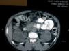
We present the case of a female patient, 41 years of age, with no significant past medical history, who was sent from the emergency department due to deterioration of renal function with serum creatinine (SCr) of 6.5mg/dl, in the setting of vaginal bleeding requiring transfusions. At that time, she was diagnosed with uterine fibroids by the gynaecology service. She was admitted for further work-up and a renal ultrasound was performed, showing grade IV bilateral hydronephrosis, with poor corticomedullary differentiation, but without visible ureters, for which a CT scan was performed. The CT showed grade IV bilateral ureterohydronephrosis secondary to extrinsic compression by the myomatous uterus, which measured 13 x 9cm (Figures 1 and 2). The urology service was notified, which placed a double “J” catheter in the right ureter, but was unable to place one in the left ureter. Evaluation by the gynaecology service was requested, which postponed a simple hysterectomy to the following week.
Following the completion of the hysterectomy, the patient had a favourable clinical progression, but not biochemically, with SCr remaining at 4.5mg/dl 15 days after surgery. Another renal ultrasound was performed, which showed grade II hydronephrosis and poor corticomedullary differentiation, therefore a percutaneous renal biopsy was not performed. The patient was discharged with the diagnosis of grade IV chronic kidney disease, secondary to probable interstitial nephritis, with follow-up in the pre-dialysis clinic. In cases of obstructive ARF, prompt resolution of the obstruction leads to complete resolution of ARF. In our case, from the time that the imaging tests were obtained, the chronicity of the process could be seen. Therefore, early diagnosis and treatment is important, since they ensure renal viability.

Figure 1. CT cut where bilateral hydronephrosis can be seen.

Figure 2. Lower CT cut where bilateral ureteral dilatation can be seen.


