Entre las complicaciones de la diálisis peritoneal, se encuentran las fugas de líquido hacia diferentes cavidades, entre ellas a la cavidad pleural. El diagnóstico de fuga diafragmática puede realizarse mediante toracocentesis o diferentes exploraciones de imagen con administración de contrastes iónicos o de gadolinio, considerándose alguna de ellas técnicas invasivas y no estando exentas de riesgo de toxicidad por contraste. Presentamos dos casos de derrame pleural en diálisis peritoneal en los que la prueba diagnóstica no invasiva mediante gammagrafía con Tc99m intraperitoneal permitió confirmar la sospecha de diagnóstica al mínimo coste. Concluimos que la gammagrafía intraperitoneal es la técnica de elección para el diagnóstico de fugas peritoneales en estos pacientes.
Pleural effusion secondary to pleuroperitoneal communication is an unusual complication of continuous ambulatory peritoneal dialysis. Many modalities have been used to diagnosis pleuroperitoneal: pleural fluid analysis, chest X- ray, Tc-99m gammagraphy, computed tomography scan and magnetic resonance image. Some of these procedures are invasive or have a high risk of induced-contrast nephrotoxicity. We present two case reports of pleuroperitoneal leak in two patients on peritoneal dialysis diagnosed with Tc-99m gammagraphy. We conclude that Tc-99m gammagraphy is a simple, safe, non invasive, low radiation exposure and cost effective method in the assessment and evaluation of complications related to peritoneal dialysis such as pleuroperitoneal leak.
INTRODUCTION
The complications associated with peritoneal dialysis include leaks affecting different cavities (pleural, retroperitoneal or inguinal). The prevalence of diaphragmatic leaks is low (2-6% of all peritoneal dialysis patients).1 They are mainly caused by direct contact between the pleura and peritoneum which is the result of damage to the diaphragmatic barrier (congenital or acquired), mainly on the right side. In the case of acquired defects, the presence of negative pleural pressure with positive intra-abdominal pressure can cause small valves to open which allow the liquid to flow in only one direction. Congenital defects, on the other hand, usually present a bidirectional flow.1,2
If a patient presents a diaphragmatic leak, his/her symptoms are similar to those of left-sided heart failure (dyspnoea, orthopnoea), and on examination he/she also presents signs compatible with the condition.
The diagnosis of a diaphragmatic leak can be made using a thoracocentesis (by detecting D-lactate isoform in peritoneal dialysis fluid, or when hyperglycaemia is present), phase contrast x-ray imaging, abdominal CT using intraperitoneal contrast and scintigraphy with intraperitoneal instillation of radioisotopes.1-5 It can also be diagnosed using a MRI and intraperitoneal administration of gadolinium contrast.6,7
The following two cases involve peritoneal dialysis patients that were admitted into hospital presenting diaphragmatic leaks. A diagnosis was made with scintigraphy using Tc99m.
CASE 1
A seventy five year-old patient with a history of high blood pressure, dyslipidaemia and stage 5 chronic kidney disease of unknown origin, was admitted into hospital to start manual intermittent peritoneal dialysis after a peritoneal catheter had been inserted. Three days after starting the treatment, the patient described shortness of breath following slight exertion. The examination of symptoms and x-ray tests were compatible with a left sided pleural effusion. Scintigraphy imaging with Tc99m confirmed the suspected leakage of peritoneal dialysis fluid into the pleural cavity. This case was managed by increasing fluid drainage and suspending peritoneal dialysis. An AVF in the left arm constructed from the humeral artery and the cephalic vein wasinserted in preparation for haemodialysis without complications.
CASE 2
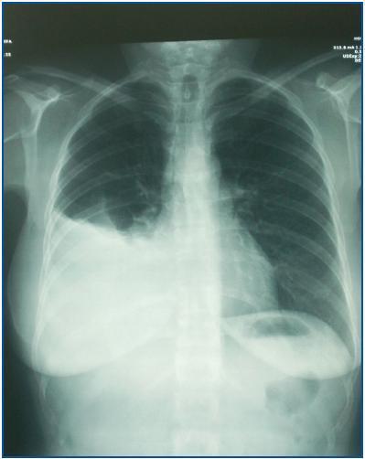
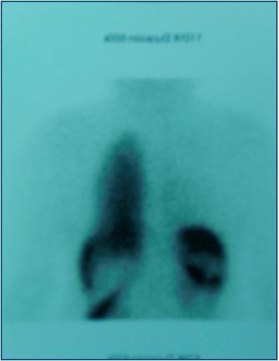
A 43 year-old patient with a history of stage 5 chronic kidney disease of unknown origin who was undergoing renal replacement therapy with manual intermittent peritoneal dialysis was admitted presenting symptoms including shortness of breath following slight exertion after starting automatic peritoneal dialysis. The symptoms were examined and x-ray tests indicated right sided pleural effusion (figure 1). The suspected diagnosis of a diaphragmatic leak was confirmed by carrying out a gamma ray imaging with Tc99m (figure 2). Peritoneal dialysis was suspended for one month and afterwards low volume, manual continuous peritoneal dialysis was initiated. The patient presented symptoms and x-ray results that were compatible with pleural effusion once again and therefore, the decision was made to suspend peritoneal dialysis and start haemodialysis using a catheter in the internal jugular vein, prior to AVF access.
DISCUSSION
Some of the aforementioned diagnostic techniques involve certain risks, including ionizing radiation and nephrotoxicity associated with the contrast agent. A diagnostic thoracocentesis is also an invasive technique. In addition, given the risk of complications associated with administering gandolinium to patients with kidney failure, and the lack of follow-up for patients in studies which have involved this technique, this cannot be considered a diagnostic technique of choice at least until more information is available regarding the potential systemic involvement of intraperitoneal gandolinium administration.
However, scintigraphy with Tc99m (2-5 mCi) is associated with a number of advantages given that it is a non-invasive technique, it is low-cost and it can provide several images without increasing the radiation that the patient is exposed to. This imaging technique begins with the intraperitoneal administration of the radioisotope. New images are taken two hours after administration (in order to blend the mixture) and then all the contrast is expelled when the peritoneal liquid is drained. The test is positive if the radioisotope is detected in the pleural cavity.2,4,5 In the aforementioned cases, a General Electric Millenium MPS gamma camera was used with 1 mCi of Tc99m, and images were acquired 5, 30 and 60 minutes after this was administered. In the cases described, less radioisotope was needed (1 mCi) in comparison with those cases found in the literature. The image quality obtained using this amount was good enough to be able to make a diagnosis.
CONCLUSION
We believe that scintigraphy and intraperitoneal Tc99m administration is the technique of choice for diagnosing diaphragmatic leaks in peritoneal dialysis patients.
Figure 1.
Figure 2.


