To the Editor,
Peritoneal infection (PI) or peritonitis is a common complication in peritoneal dialysis (PD). It can damage the peritoneal membrane, compromise the technique’s survival rate and is the main cause for transfer to haemodialysis.1 Most PI episodes are due to gram-positive cocci, although an increase in PI due to gram-negative bacilli is being observed, with worse clinical prognosis.2
Oerskovia infections are rare in humans. There are only four oerskovia PI cases published.3-6 It consists of two large species: Oerskovia xanthineolytica (O. xanthineolytica) and Oerskovia turbata (O. turbata), which are currently known as Cellulosimicrobium cellulans7 and Cellulosimicrobium funkei, respectively,8 according to comparative phylogenetic analysis based on 16S ribosomal rRNA gene sequences. They are gram-positive bacilli found in the ground, pastures and water from different parts of the world. Colonies are characterised by a yellow pigment with branching vegetative hyphae which penetrate in the agar.9,10
Below, we describe the first peritonitis case caused by O. turbata in a patient undergoing automated peritoneal dialysis (APD), and have also reviewed published cases.
We present the case of a 62-year-old man, with a history of high blood pressure, dyslipaemia, stroke and ischaemic heart disease. He started APD in August 2006 following bilateral kidney nephrectomy for transitional cell carcinoma. He presented with his first Pasteurella multocida PI in October 2008, which was presumably induced by contact with a cat.
In July 2009, he had his second PI. He started drug treatment with vancomycin, intraperitoneal gentamicin, and ciprofloxacin orally. On the third day, gram-positive bacilli growth was found with a preliminary identification of Oerskovia sp. On the sixth day, the antibiogram showed an intermediate sensitivity to ciprofloxacin and sensitivity to co-trimoxazole. Ciprofloxacin was then replaced with oral co-trimoxazole, intraperitoneal vancomycin regimen was continued every 5 days, and gentamicin was withdrawn. On the tenth day, peritoneal fluid was clear. Antibiotic treatment was maintained for 22 days.
Thirteen days after finishing the antibiotic treatment, the patient had a PI relapse. Treatment using vancomycin, intraperitoneal gentamicin and oral co-trimoxazole was restarted, using fluconazole as prophylaxis. On the third day, fluid was clear and Oerskovia sp growth was resistant to co-trimoxazole, and sensitive to ciprofloxacin. The antibiotic regimen was therefore changed: gentamicin was withdrawn and vancomycin maintained. Twenty-one days after antibiotic treatment, he had a second PI relapse. The PD was removed and the patient was transferred to haemodialysis. Catheter culture was negative.
Microbiology
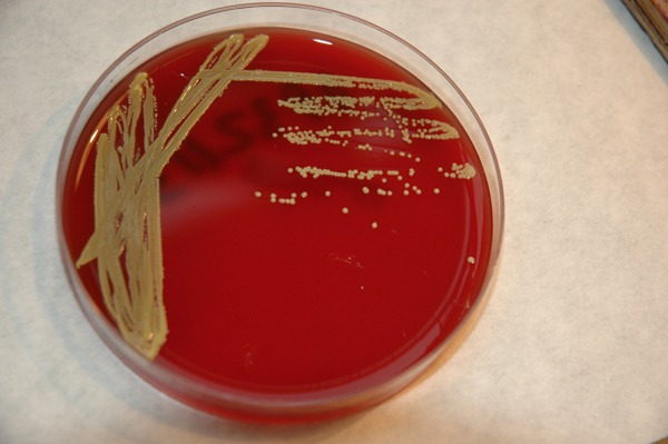
The peritoneal fluids were collected in aerobic and anaerobic bottles for BacT/Alert blood culture test (bioMérieux, Durham, NC, USA). Both bottles were found to be positive at between 23 and 24 hours, and the gram staining revealed branching gram-positive bacilli. Chocolate agar, blood agar, and colistin-nalidixic acid (CNA) agar were reseeded on blood agar plates, which were incubated at 35ºC in 5% CO. Some colony growth was observed at 48-72 hours. It was the characteristic pastel yellow colour (Figure 1). The colonies were catalase-positive and oxidase-negative, and were identified using API Coryne galleries (bioMérieux, Marcy L’Étoile, France). It had the code 7572727, with an Oerskovia sp. percentage of 99.9%. Later, we completed the 16S rRNA sequencing, and the phylogenetic gene sequence analysis confirmed Cellulosimicrobium funkei.
Review of the PI due to Oerskovia cases
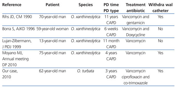
We have found four cases of PI due to O. xanthineolytica PD in the literature. Catheter removal was necessary in two of these cases.3,6 Cases with good progress were treated with intraperitoneal antibiotics for 15 days4,5 (Table 1).
We would like to present the first PI due to O. turbata. This species is less frequent in human infections than O. xanthineolytica according to the review published in 2006.7 This is the first case of infection caused by Oerskovia diagnosed in our health centre. At present, according to the comparative phylogenetic analysis based on 16S rRNA gene sequence, it is called Cellulosimicrobium.
Human infections due to Oerskovia develop in immunosuppressed patients and foreign body carriers: central catheters, valvular and joint prostheses, and peritoneal dialysis catheters.3-7,9,11 In some cases, the origin of the infection has been related to working or living in rural areas.5,10
More than one antibiotic may be necessary and the foreign body may need to be removed. In all cases the germ has been sensitive to the antibiotic treatment in in vitro tests, although in vivo results have been poor.7 Our case, despite correct antibiotic treatment, did not manage to eradicate the infection and the PD catheter had to be removed.
This bacterium is resistant, although not especially virulent, given that no deaths have been recorded.7,12 Most infections caused by O. turbata have been in immunosuppressed patients.13-15 Our patient lived in an urban area and had a pet cat, but there are no references that show a relationship between infections caused by Oerskovia and domestic animals.
The Oerskovia genus is often in vitro resistant to penicillin, aminoglycosides, macrolides and cephalosporins, and intermediate resistance to ciprofloxacin. It is considered in vitro sensitive to vancomycin and rifampicin.7,12 In this instance, the germ was sensitive to vancomycin, rifampicin, meropenem and co-trimoxazole, and was intermediate sensitive to ciprofloxacin. Given that there are no standardised minimal inhibitory concentration (MIC) values to study the sensitivity of this bacterium, we used the MIC for Corynebacterium sp.
To conclude, PIs caused by Oerskovia are rare despite it being a germ that is widely distributed in nature. It can affect immunosuppressed patients and immunocompetent foreign body carriers. Response to antibiotic treatment is often poor and catheter removal may be required. This bacterium may be more common in humans in the future, due to its distribution in nature, and the increased number of immunosuppressed patients with prosthetic material.
Figure 1. Oerskovia culture on a blood agar plate
Table 1. Review of cases of PI caused by Oerskovia


